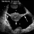
Figure 15.1.1
Definite pregnancy failure. Transvaginal sonogram demonstrates an intrauterine gestational sac with an embryo (calipers) whose crown-rump length measures 5.33 mm. No cardiac activity was seen on real-time sonography.
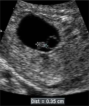
Figure 15.1.2
Probable pregnancy failure. Transvaginal sonogram demonstrates an intrauterine gestational sac with an embryo (calipers) whose crown-rump length measures 3.5 mm. No cardiac activity was seen on real-time sonography.
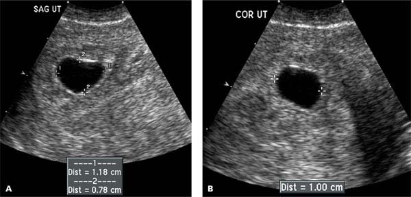
Figure 15.1.3
Probable pregnancy failure. (A) Coronal and (B) sagittal views through the uterus demonstrate an intrauterine gestational sac (calipers) whose mean sac diameter is 9.9 mm (average of 11.8, 7.8, and 10.0 mm). Because no yolk is seen and the mean sac diameter is >8 mm, the likelihood of pregnancy failure is high.
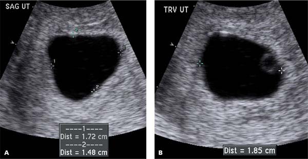
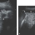
Figure 15.1.4
Probable pregnancy failure. (A) Sagittal and (B) transverse views through the uterus demonstrate an intrauterine gestational sac (calipers) whose mean sac diameter is 16.8 mm (average of 17.2, 14.8, and 18.5 mm), containing a yolk sac. Because no embryo is seen and the mean sac diameter is >16 mm, the likelihood of pregnancy failure is high.
15.2. Subchorionic Hematoma
Description and Clinical Features
Many women experience vaginal bleeding during the first trimester of pregnancy. Sometimes, in addition to blood passing per vagina, blood collects between the chorion of the developing gestation and the uterine wall, forming a subchorionic hematoma. When a hematoma is present, the pregnancy prognosis is related to the size of the hematoma. The prognosis is excellent if the hematoma is small, whereas large hematomas are associated with a fairly high risk of pregnancy loss.
Sonography
On ultrasound, a subchorionic hematoma appears as a hypoechoic or anechoic area around part of the gestational sac, outside the chorion (Figure 15.2.1). It is often crescentic in shape. A subchorionic hematoma must be distinguished from chorionic fluid, which is located between the chorion and amnion and is a normal finding in the first trimester. With a subchorionic hematoma, there are two fluid collections—the hematoma and the fluid within the gestational sac—that are separated by the relatively thick chorion (Figure 15.2.1). Chorionic fluid, on the other hand, is separated from amniotic fluid by the thin, smooth amnion (Figure 15.2.2).
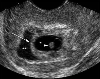
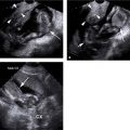
Figure 15.2.1
Subchorionic hematoma. Transverse view of the uterus demonstrates two fluid collections: an inner collection (*) of fluid within the gestational sac, and a surrounding crescentic fluid collection (**), representing a subchorionic hematoma. These two fluid collections are separated by the thick chorion (arrow). The embryo (arrowhead) is visible within the gestational sac.
Related posts:


Stay updated, free articles. Join our Telegram channel

Full access? Get Clinical Tree


