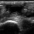
Fig. 18.1
Glass fragment. Sonography shows a hyperechoic fragment with posterior reverberation (arrow) in the subcutaneous tissue. Posterior reverberation artifacts are characteristic of glass fragments
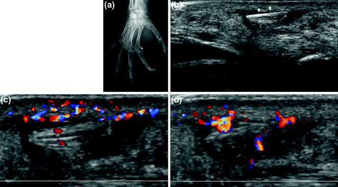
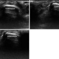
Fig. 18.2
Metal sliver. Radiography reveals a small sliver near the second metacarpal head. The intense radiopacity of the object is typical of metal (a). On sonography, the sliver appears hyperechoic (arrows) (b) with posterior reverberation and a halo of granulation tissue, which appears hypoechoic and hypervascularized (c, d)
Table 18.1
Ultrasonographic appearance of the foreign bodies most commonly found in the hand and wrist
Glass fragments | Hyperechoic objects with posterior reverberation artifacts |
Metal fragments | Hyperechoic objects with posterior attenuation |
Wooden splinters, thorns, cactus spines | Hyperechoic objects with no reverberation or posterior artifacts |
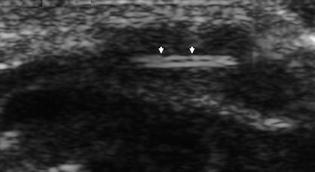
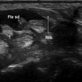
Fig. 18.3
Wood splinter. The sonographic examination reveals a hyperechogenic object (arrows) with no attenuation or posterior reverberation. Like many splinters, this one is surrounded by hypoechoic granulation tissue
While radiography can easily identify the presence of a radiopaque foreign bodies [1, 2], sonography can also reveal those that are radiolucent (cactus spines, thorns, wood splinters, and fragments of nonradiopaque glass) [1, 2, 4, 5].
Related posts:
Stay updated, free articles. Join our Telegram channel

Full access? Get Clinical Tree


