Score is a total of 3 components. Score ≤ 3 suggests nonoperative treatment, while score of 4 is indeterminate. Score ≥ 5 suggests operative treatment. For injury mechanism, the worst level is used, and the injury is additive. An example is distraction injury with burst without angulation is 1 (simple compression) + 1 (burst) + 4 (distraction) = 6 points. (Vaccaro 2006.)
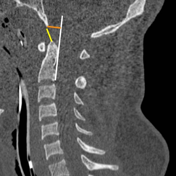
(Left) Sagittal graphic shows normal basion-dental interval (BDI) (red line) and the basion-posterior axial line interval (BAI) (yellow line). BAI is the distance from basion to posterior axial line (black line). BDI is abnormal if > 10 mm on sagittal CT. BAI is abnormal if > 12 mm on plain films.
(Right) Sagittal NECT of trauma patient with AOD shows abnormal distance between basion and dens (yellow), abnormal separation of basion from posterior axial line (orange). Posterior axial reference line is white.
(Left) Parasagittal NECT in a patient with AOD shows widening of both C0-C1 and C1-C2 articulations . Summed condylar displacement (sum of the bilateral distances between midpoint of occipital condyle and C1 condylar fossa) is abnormal if > 4.2 mm.
(Right) Sagittal STIR shows abnormally widened and hyperintense C0-C1 and C1-C2 articulations . This patient underwent occiput to C3 posterior fusion for atlantooccipital and atlantoaxial dislocation.
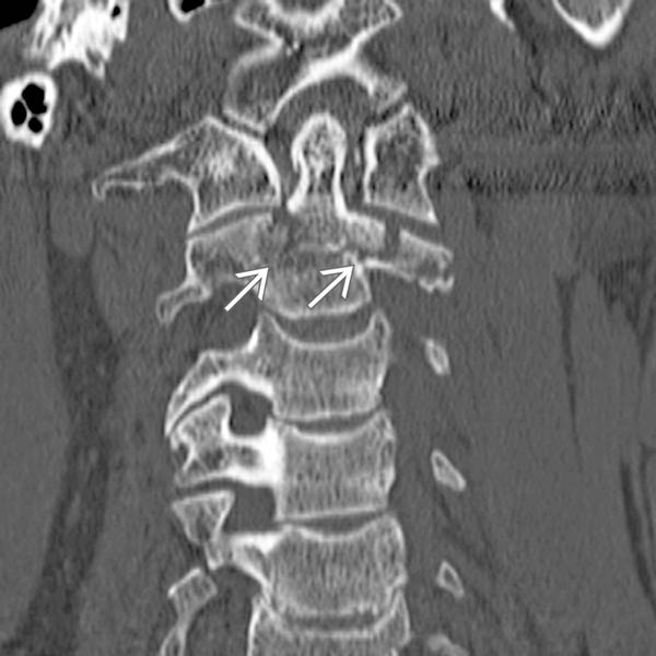
(Left) Axial NECT shows multiple fracture sites involving the C1 ring without canal compromise.
(Right) Axial NECT shows fractures involving both anterior and posterior rings of C1 and additional avulsion fracture off of mesial C1 ring at the level of attachment of transverse ligament .
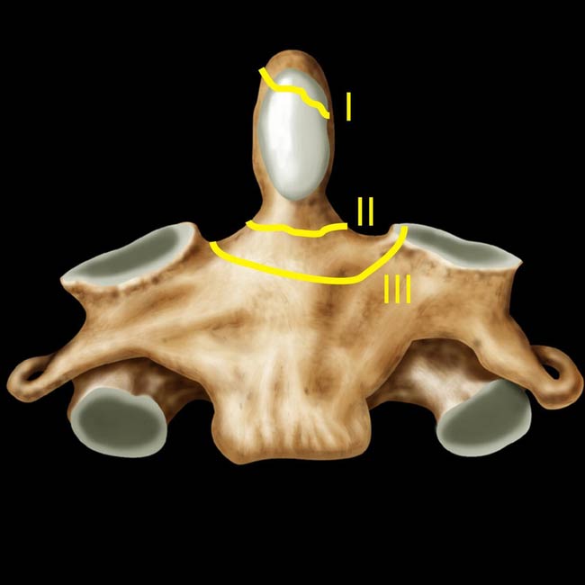
(Left) Coronal graphic of the C2 vertebra shows the schematic for location of types I, II, and III odontoid fractures.
(Right) Coronal NECT shows an oblique type III fracture extending across the base of odontoid and the upper body of C2 with fragmentation of the left lateral mass of C2.
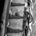
(Left) Sagittal NECT shows a C2 pars fracture without significant offset, angulation, or distraction.
Only gold members can continue reading. Log In or Register to continue

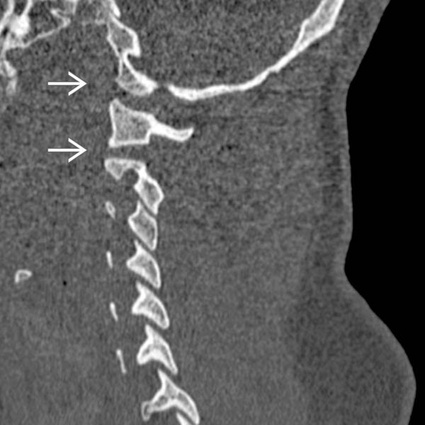
 . Summed condylar displacement (sum of the bilateral distances between midpoint of occipital condyle and C1 condylar fossa) is abnormal if > 4.2 mm.
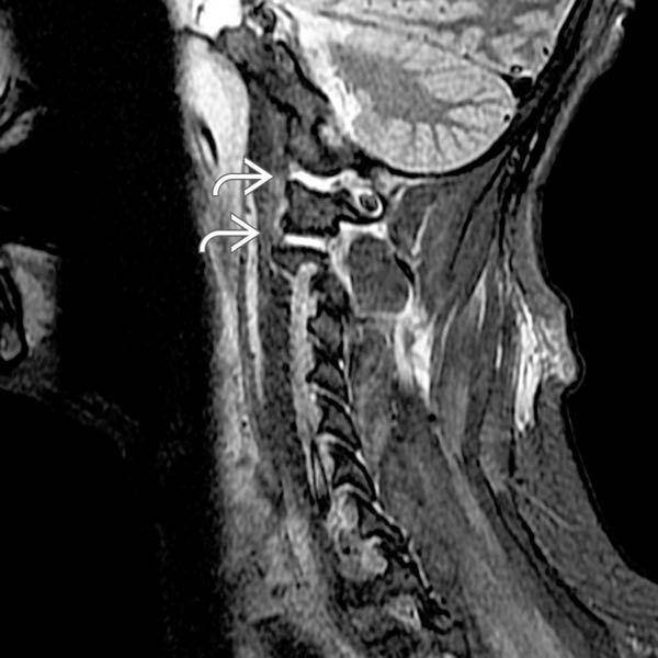
 . This patient underwent occiput to C3 posterior fusion for atlantooccipital and atlantoaxial dislocation.
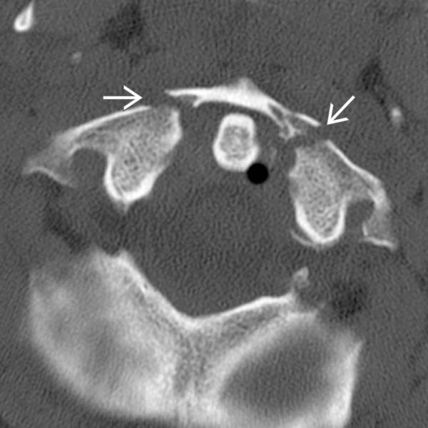
 without canal compromise.
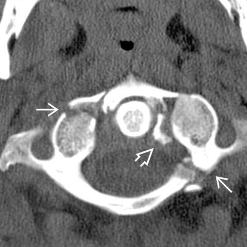
 and additional avulsion fracture off of mesial C1 ring at the level of attachment of transverse ligament
  .
.
 with fragmentation of the left lateral mass of C2.
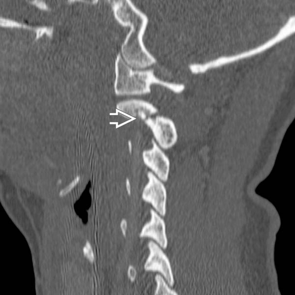
 without significant offset, angulation, or distraction.