© Springer International Publishing Switzerland 2015
Patricia M. Carrascosa, Ricardo C. Cury, Mario J. García and Jonathon A. Leipsic (eds.)Dual-Energy CT in Cardiovascular Imaging10.1007/978-3-319-21227-2_1515. Future in Dual Energy CT
(1)
Division of Cardiology, University of Washington, 1959 NE Pacific Street, 356422, Seattle, WA 98195-6422, USA
Abstract
Dual energy CT has progressed rapidly over the past few years and is making inroads into routine clinical care for a spectrum of diseases. In the field of cardiovascular medicine, dual-energy CT has been applied to the evaluation of myocardial ischemia, myocardial viability and atherosclerotic plaque characterization.
However, most of these applications remain in the research realm at present and further advancements are needed.
The future of dual-energy CT likely lies in some modifications to overcome current limitations of high radiation dose and artifacts with ability to provide enhanced visualization and improved temporal resolution.
DECT has seen relatively rapid advances and new applications mainly in relation to hardware and software improvements that have enabled improved image quality. DECT, supported by rapid advancements in spatial and temporal resolution plays an active role in a number of cardiovascular imaging applications. Nonetheless DECT has much maturation to undertake to achieve its ultimate goal of true spectral imaging and tissue characterization.
Keywords
Future dual energy CTPhoton countingTemporal resolutionDual energy CT has progressed rapidly over the past few years and is making inroads into routine clinical care for a spectrum of diseases. In the field of cardiovascular medicine, dual-energy CT has been applied to the evaluation of myocardial ischemia [1], myocardial viability [2], and atherosclerotic plaque characterization [3] as outlined in the previous chapters. We have also seen significant investigation and validation of reduced contrast CT angiography supported by the ability to increase signal in the vascular bed through low monochromatic energy imaging [4, 5]. However, most of these applications remain in the research realm at present and further advancements are needed.
It is interesting to note that the future of CT is firmly rooted in the past. Enhanced discrimination and thus higher spatial resolution and tissue characterization with dual-energy CT was actually first described by Dr. Hounsfield in 1973. Similarly, the photon counting CT scanner, where the number as well as the energy of the incoming photons are measured, was conceived over 30 years ago. Further, many of the difficulties identified in the early development of CT, such as complex image processing, basis material decomposition, temporal speed, artifacts, and photon discrimination, still exist albeit to a lesser extent. With recent software, hardware and technological advances, dual-energy or spectral CT is poised to fundamentally change cardiac CT scanning and image evaluation.
Future of Hardware Advancements
Dual Energy CT
As has been described in previous chapters, there are currently three dual-energy CT systems in clinical use. Their goal, as with any future hardware advancement in CT is aimed towards faster production of image with fewer artifacts and radiation dose while enhancing visualization and tissue characterization. The dual-source dual-energy system of Siemens has a significant high energy separation and a temporal resolution advantage, especially when the high pitch mode can be used. However, image co-registration is challenging given the 90° difference in tube angle and the resulting temporal skew of data [6]. On the other hand, since the single-source dual-energy fast kVp switching system of General Electric can simultaneously acquire data between low and high energies at less than quarter of millisecond apart, it has the advantage of better image co-registration while acquisition speed and in plane energy differentiation is more limited [7].
A single-source dual-energy scanner with dual detector layers of Phillips is another example of advancement in CT hardware for dual-energy CT which could allow simultaneous high and low energy discrimination [8].
Large coverage detector may allow to scan the entire cardiac cycle of the heart in one gantry rotation and perform perfusion assessment with similar contrast enhancement in anterior and inferior segments. The future of dual-energy CT likely lies in some combination of the latter modifications to overcome current limitations of high radiation dose and artifacts with ability to provide enhanced visualization and improved temporal resolution.
Photon Counting and Spectral CT
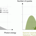
In conventional CT systems, scintillation-based detectors measure the photon intensity after the x-ray beams interact with tissue by either Compton scatter or photo-electric effects. Whereas dual energy has the potential to differentiate and capitalize on these two effects to create monochromatic images for different tissue components, important data are lost. The interactions of photons and tissue not only affect the number of photons passing through the body part but also the photon energy that reach the detector. Detectors that can measure different energy spectra, such as the current dual layer detector by Philips, differentiate not only the incident photons but photon energy as well [9] More advanced “photon-counting” detectors simultaneously measure the number of incident photons, as well as discriminate and tabulate the different photon energies. The number as well as the relative energy of photons can then be placed in energy “bins” which allow creation of spectral energy distributions As opposed to the “source-based” spectral energy delivered with dual energy CT, “detector-based” CT measurements with photon counting detectors along the spectrum of photon energies allow for more reined material decomposition than dual-energy CT alone.
There are many advantages to spectral CT imaging. First, image noise can be decreased by the direct conversion of photon energy into electronic measures which, in turn, lower electronic image noise [10]. This would translate to better image quality with similar or lower radiation doses than today. Secondly and more importantly, spectral measurements from each x-ray beam can both qualitatively and quantitatively differentiate human tissues and injected contrast agents more accurately than current scanners. Using K-edge or other imaging techniques, more than two measurements for each energy level can enhance quantitative and molecular imaging. Spectral CT has the potential to better differentiate tissue types important in cardiovascular disease, including stable, unstable, or calcified atherosclerotic plaque, myocardial fibrosis, and iodinated contrast concentrations. Further, multiple contrast agents injected simultaneously could be differentiated for molecular imaging applications.
The challenges to development of spectral CT are daunting and have delayed its emergence into clinical medicine for over 30 years. Most current detectors are scintillator-based converting x-ray photons into optical photons which are detected by a photodiode; this binary system does not allow for photon energy discrimination. Further, since detection of small changes in the K-edge are needed for energy discrimination, newer detectors, such as the CdTe semiconductor based detector, x-ray photons are converted into electron hole pairs that can be discriminated into individual photon number and energy. The measured number of electron hole pairs are proportional to x-ray photon energy and are used to separate the different binned photon energies into quantity (photon counting) and spectral energy data. In addition, spectral CT detectors are relatively inefficient for iodine-containing contrast concentrations due to the low K-edge of iodine (keV = 33). To take full advantage of K-edge imaging, contrast with higher attenuation due to higher anatomic number such as gadolinium or metals, as gold (81 keV), will need to be used. Other agents are not currently available in clinical care and will have to be optimized for signal as well as for toxicity of the agent [11]. Given all the potential benefits, photon-counting and spectral CT appear likely to be the next true revolution in CT imaging.
Future of Software Advancement
Parallel rapid evolution of computational technology and software development has complimented CT hardware advancements extremely well. Filtered back projection is the most commonly used reconstruction algorithm in use for CT as it is fast and robust though limited by poor image quality especially in low radiation-dose acquisition protocols or in obese patients [12]. Since lowering radiation dose is paramount to the future of CT applications, advance iterative reconstruction algorithms which were utilized in early days of CT are being reutilized more effectively with current computational technology [13]. While they are still being developed, current iterations have shown the potential to augment spatial resolution and material separation of dual-energy CT by to reducing image noise three to tenfold [14, 15]. Current software includes adaptive iterative reconstruction, such as Adaptive Statistical Iterative Reconstruction V (ASIR V) General Electric, SAFFIRE of Siemens, Adaptive Iterative Dose Reduction 3D (AIDR) of Toshiba and iDose/IMR of Phillips.
In some approaches such as the kpv switching IR is available until 60 kev. It will be possible in the near future to use it up-to 40 kev, in this way improving image noise.
Stay updated, free articles. Join our Telegram channel

Full access? Get Clinical Tree


