Features
SOMATOM definition DS
SOMATOM definition flash
SOMATOM force
Discovery 750HD
Revolution
Brilliance iCT SP 128
Brilliance iCT 256
Aquilion one
Vendor
Siemens
Siemens
Siemens
GE
GE
Philips
Philips
Toshiba
Technique used for dual energy
Dual source
Dual source
Dual source
Fast kVp switching
Fast kVp switching
Singe source dual scan
Singe source dual scan
Single source dual scan
Number of detectors
2
2
2
1
1
1
1
1
Number of detector elements (z-axis)
32
64
96
64
256
64
128
320
Detector configuration
2 × 32 ×0.6
2 × 64 × 0.6
2 × 96 × 0.5
64 × 0.625
256 × 0.0625
64 × 0.625
128 × 0.625
320 × 0.5
Detector width (mm)
19.2
38.4
48
40
160
40
80
160
Number of slices
2 × 64 (128)
2 × 128 (256)
2 × 192 (384)
2 × 64 (128)
2 × 256 (512)
2 × 64 (128)
2 × 128 (256)
320
Slice thickness [mm]
0.6/1.2
0.6/1.2
0.5/1
0.625/1.25
0.625/1.25
0.625/1
0.625/1
0.5/1
Field of view (FOV) [cm]
26a
33a
50b
50
50
50
50
50
Gantry rotation time for dual energy acquisition [ms]
330
300
258
350
280c
800
800
550
Temporal resolution for dual energy acquisition [ms]
165
150
129
175
140
400
400
275
Dual Source Dual-Energy CT
Dual-source CT contains two sets of x-ray tube and detector arrays (two X-ray tubes and corresponding detectors), which are arranged in a single gantry perpendicular or almost perpendicular (90–95°) to each other in the x-y plane (Fig. 10.1). With two x-ray sources, a DSCT system allows two different kV levels and mAs settings to be used simultaneously (Fig. 10.2).
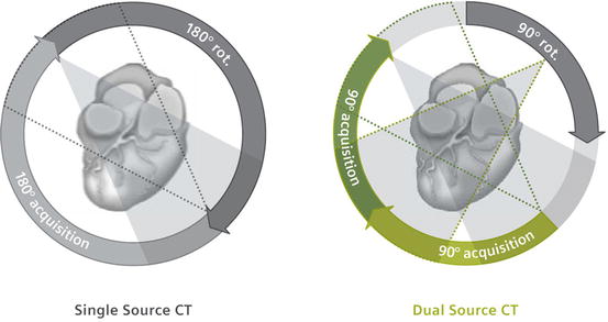
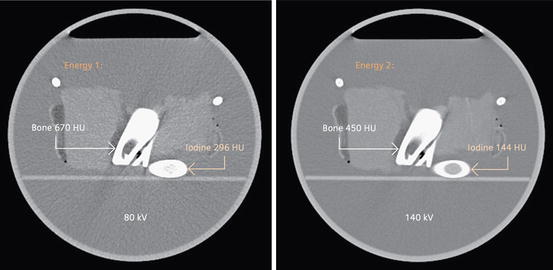
Fig. 10.1
Schematic illustration of the principle of dual energy imaging with a DSCT system. Both tube/detector systems are mounted with an angle of 90°; images at low and high voltage are acquired simultaneously (Image courtesy of www.healthcare.siemens.com)
Fig. 10.2
Because x-ray absorption is energy dependent, changing the tube’s kilovoltage results in a material-specific change of attenuation (Image courtesy of www.healthcare.siemens.com)
Dual-source CT has several advantages over the single-source scanners, depending on the mode of scan acquisition. First, both X-ray tubes can be operated simultaneously in a standard spiral or sequential acquisition mode and the tube voltage and current can be adjusted freely to obtain the largest possible difference in photon energies with similar total amounts of quanta from both tubes. Second, imaging the same slice with the two tubes on the same time improve temporal resolution and allows for robust cardiac imaging. One-quarter rotation time is sufficient to reconstruct images without the need for a multisegmental reconstruction technique. By using the two tubes at identical kVp levels, it is possible to acquire images using data from only 90° of gantry rotation instead of the conventional 180° of data required for the single-source CT. The resultant temporal resolution of only 83 ms (doubling the temporal resolution) is particularly advantageous in coronary artery imaging. This makes it possible to obtain diagnostic images at higher heart rates than previously possible and possibly without the need for β-adrenergic blockers. Additionally, both tubes can be used to increase photon flux that offers a diagnostic benefit in morbidly obese patients. Operating the two tubes at different tube potentials enable simultaneously acquisition of two scans with potential applications for tissue differentiation. This would make further material differentiation possible compared with that achieved with x-ray attenuation in Hounsfield units. That facilitates classification of different tissue types and furthers helps in the characterization and differentiation of different types of atherosclerotic plaque, e.g. calcified and non-calcified plaque that will help to improve risk stratification of cardiovascular patients.
With the first two generations of dual source MDCT scanners, the space in the CT gantry was only sufficient for a smaller second detector so that the field of view of the dual energy scans was restricted to 26 cm for the first generation system (SOMATOM Definition DS) and 33 cm for the second generation system (SOMATOM Definition Flash). This has been overcome in the latest generation scanner, the SOMATOM Force where the field of view for dual energy CT is now 50 cm. There also concern in dual systems regarding the susceptibility of the system to cardiac motion due to the time difference between data acquisition of the two tubes as well as the possibility of image degradation due to cross-scatter from each tube. A final disadvantage of this approach is the increased cost by adding a new hardware, making the system more expensive.
Currently, there are three clinical, commercially available Dual Source CT (DSCT) Scanners: the SOMATOM Definition, the SOMATOM Definition Flash and the SOMATOM Force (all by Siemens). For dual energy scan modes, the SOMATOM Definition is operated at 80 kV/140 kV, while the SOMATOM Definition Flash has an additional tin filter (Sn) and is typically used at 100 kV/Sn140 kV. The Force can be used in 10 kV increments ranging between 70 and 150 kV allowing for high-quality DE scans. At low kV, high mAs must be used (up to 900 mAs) which has been one of the rate limiting steps to CT data acquisition at lower kV energies.
Rapid Voltage Switching
Dual-energy scanning can also be performed using single-source detector units by using a rapid voltage switching technique (Fig. 10.3). This is achieved by using projection data acquired in both axial and helical modes, unlike the image-based dual-energy processing of dual-source CT. The fundamental principle behind fast kVp switching is the acquisition of two different datasets with the source voltage alternating between low and high kVp in a single rotation. This enables precise temporal registration of views, thereby freezing motion as the alternating spectrums penetrate the patient. The generator and tube are capable of reliably and rapidly switching between 80 and 140 kVp targets, and have the capability to support sampling as quickly as every 150 μs with less than 0.25 ms required to switch between the two energies in the latest generation scanner. The Gemstone detector is a key contributor to fast kVp switching acquisitions through its scintillator and data acquisition system; this will be discussed later in this chapter. This solution has the major advantage of decreased cost, because there is no need to equip the unit with additional hardware.
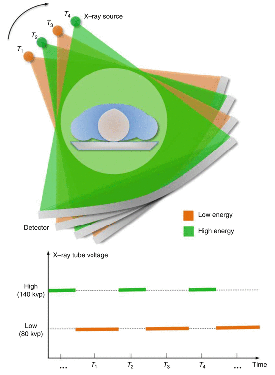
Fig. 10.3
Rapid voltage switching (‘fast kV switching’) as utilized in the GE dual-energy systems (Image courtesy of GE Healthcare)
The disadvantages of single-source dual-energy scanning are the unequal noise levels that may result in the datasets owing to the rapid modulation of tube current and the fact that the use of the same filtration for the two energy beams may result in suboptimal spectral separation. Additionally, the rotation speed of the system may have to be reduced to account for the acquisition of these additional projections and the rise and fall times of the voltage modulation that prolongs the acquisition time and decreases temporal resolution.
Spectral CT and Dual-Layer Detectors
Philips Healthcare makes a distinction between “spectral CT” and “dual-energy CT”, terms that have traditionally been used almost interchangeably in the past. Philips’ newest addition is the IQon Spectral CT, and based on the company’s NanoPanel Prism design. This uses a spectral detector that detects X-ray photons of more than one energy simultaneously. Designed to become a new class of CT where high and low energies are sampled in the same time and space that technically allows for conventional anatomical information to be obtained at the same time as material characterization and monoenergetic image information in one scan. This may obviate the need for radiologists to determine what clinical information is needed at the time of the scan, as the IQon system will obtain all of the information that can be accessed at any time. The company likens this technology to a prism, which can split a beam of white light into the colors of the rainbow. Similar to other manufacturers, the spectral CT system also has the ability to perform virtual non-contrast CT, thus reducing the need for an initial non-contrast CT in multiphase examinations. For example, the system can identify iodine and calcium and may be useful in determining kidney stones and characterizing plaques. Reduction of iodinated contrast dose, less beam hardening artifact and specific lesion characterization are further benefits that are expected with this new technology.
Spectral CT enables the identification of specific element K-edges, which enables imaging of metals using CT. This may allow iron, gold or other compounds to become viable next generation CT contrast agents as metals and other elements can be combined into tailored nanoparticles in sizes between 1 and 100 μ and in different configurations to target specific receptors. The company hopes to validate IQon spectral CT for entering specific atomic numbers off the periodic table of elements to filter images to show concentration of these elements in the body which may allow new ways to characterize tissue or plaque composition based on its chemical makeup.
The detector is composed of two layers of different scintillator material, one placed (or ‘sandwiched’) directly on top of the other, in conjunction with a single x-ray tube operated at a peak tube potentials ranging from 80 to 120 kVp. The top layer is 1 mm of ZnSe (Zinc Selenide) and the bottom layer is 2 mm of GOS (Gadolinium orthosilicate). Low- and high-energy data sets are produced as the top layer absorbs photons in the low energy portion of the spectrum and the bottom layer absorbs the remaining higher energy photons. Perfect spatial and temporal registration of data sets is obtained as there is only one x-ray source and dual energy separation happens at detectors arranged one on top of the other. This eliminates X-ray dead time during acquisition making it ideal for imaging moving targets. The first version of the spectral detector CT scanner (Brilliance 64) provided 40 mm of coverage with 64 × 0.625 mm collimation, the most recent generation scanner provides 80 mm of coverage with 128 × 0.625 mm collimation. Gantry rotation speeds are as fast as 270 ms.
Projection space decomposition is used to first solve the individual Photoelectric and Compton basis components followed by reconstruction of the images. Projection based decomposition requires spatial and temporal alignment, which is achieved with the dual layer detector. Material make up is known at each sample which permits more accurate beam hardening correction, a potential advantage of projection space decomposition compared to image space decomposition. This enables the reconstruction of virtual monochromatic images with accurate beam hardening correction. It also enables reconstruction of specific material basis pairs (e.g. iodine and water), material specific and effective atomic number images.
Disadvantages of this system include the lack of ability to vary the x-ray beam filtration at the source that could confound spectral separation as it uses a single system-ray tube.
Also, noise levels are not equivalent, as the same tube current must be used during data acquisition of both the high and low energy data sets. Finally, there is a fixed energy partition between layers so only one low/high peak tube voltage combination is possible.
Several types of images can be reconstructed via dual-layer scanning technology depending on the clinical requirement. Conventional CT images using standard filtered back-projection or iterative reconstruction techniques can be generated and images are equivalent to a conventional CT image obtained from a conventional scanner with poly-energetic X-ray spectra. Virtual monochromatic images from 30 to 200 keV can be reconstructed as well as material decomposition and effective atomic number based images. These are well described in the dedicated chapter by Rajih and Halliburton (Technical Aspects of DECT with Dual Layer Detectors). The IQon scanner is currently pending FDA clearance and at the time of writing has not been cleared for sale in the USA or Canada.
Related posts:

Stay updated, free articles. Join our Telegram channel

Full access? Get Clinical Tree


