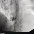
Douglas O. Faigel1 and Sarah A. Rodriguez2 1 Mayo Clinic College of Medicine, Scottsdale, AZ, USA 2 Oregon Health and Science University, Portland, OR, USA Endoscopic ultrasound (EUS) is frequently employed for the diagnosis and staging of gastric cancers and lymphomas. This chapter reviews EUS imaging and staging of these malignancies. The majority of gastric malignancies are adenocarcinomas. They are staged using the American Joint Commission on Cancer Staging TNM Classification (Table 13.1). These cancers may be either of the intestinal or diffuse type. The intestinal type arises from the mucosa and form discrete tumors. They appear as poorly circumscribed hypoechoic lesions which, at the edges, can be seen to be arising from the mucosal layers. T1 lesions are limited to the mucosa (first and second layers) or may penetrate into the submucosa (third layer) (Figure 13.1). There should be a demonstrable, intact, bright layer of submucosa between the lesion and the dark band of the muscularis propria (fourth layer). T2 lesions extend into but not through the muscularis propria (Figure 13.2). Endosonographically, T2 lesions extend through the bright third layer corresponding to where the tumor penetrates through the submucosa into the muscularis propria. However, the interface at the outer margin of the muscularis where it contacts the serosa (fourth and fifth layers) is smooth and undisturbed by the cancer. In T3 lesions the hypoechoic lesion extends completely through the fourth layer, and the serosa (fifth layer), which would otherwise be smooth, is interrupted and clearly invaded (Figure 13.3). Finger‐like projections of tumor, termed pseudopodia, may be seen extending into the extragastric space. If the lesion extends into a local organ (e.g. liver, pancreas, spleen, diaphragm) or large vessel (e.g. aorta, celiac axis) it is classified as a T4‐stage lesion (Figure 13.4). Diffuse‐type adenocarcinoma (also known as “linitis plastica”) are poorly differentiated tumors that infiltrate the stomach wall. Histologically, they consist of single cells or small clusters of cells that contain large mucin vacuoles pushing the nucleus to one side to produce a signet ring appearance. The result of the diffuse infiltration by the cancer cells is a thickened, rigid stomach that has been likened to a leather bottle. Endoscopic ultrasound examination has been found to be exceptionally helpful in evaluating the patient with a suspected infiltrating malignancy. The normal stomach is 3–4 mm in thickness. When an infiltrating cancer is present, the stomach is thickened to greater than 4 mm, and one of two EUS patterns may be seen. In the first, there is complete loss of the normal five‐layer pattern, with the markedly thickened wall assuming a homogeneously dark appearance. All layers of the stomach are generally involved and these tumors are stage T3. In the second EUS pattern, the thickened stomach maintains its five‐layer pattern, but the muscularis propria (fourth layer) is a prominent, thick, dark band beneath a thickened, bright, third layer (submucosa) (Figure 13.5). Finding either of these patterns should prompt an aggressive search for an infiltrating malignancy. However, this finding is not specific to gastric adenocarcinoma and may be seen in metastatic cancer to the stomach (Figure 13.6) and in gastric lymphoma. The stomach is the most common site for primary extranodal lymphoma, comprising one‐fourth of all extranodal cases and at least half of all primary gastrointestinal (GI) lymphomas. Endoscopically, primary gastric lymphomas usually appear as an exophytic mass, although a more diffuse infiltration can occur, causing a linitis plastica appearance (Figures 13.7 and 13.8). Gastrointestinal lymphomas are staged differently than carcinomas (Table 13.2). Tumors confined to the GI tract are stage IE, and these patients have significantly higher survival rates than those with lymph node involvement or distant spread. Table 13.1 American Joint Committee on Cancer (AJCC) Staging: TNM classification for gastric cancer (with permission from AJCC).
13
Gastric Cancer
Tumor (T) stage
Tx
Primary tumor cannot be assessed Related posts:
![]()
Stay updated, free articles. Join our Telegram channel

Full access? Get Clinical Tree

 Get Clinical Tree app for offline access