and Filiz Özülker1
(1)
Nuclear Medicine, Okmeydani Training and Research Hospital, Istanbul, Turkey
12.1 Case 1: Gastrointestinal Stromal Tumor Progression
History
A 33-year-old male status post biopsy from a mass lesion in abdomen was diagnosed to have gastrointestinal stromal tumor (GIST) 5 months ago. The patient has been treated with imatinib mesylate (Gleevec) and referred to 18F-FDG PET/CT scan for evaluation of therapy response.
Findings
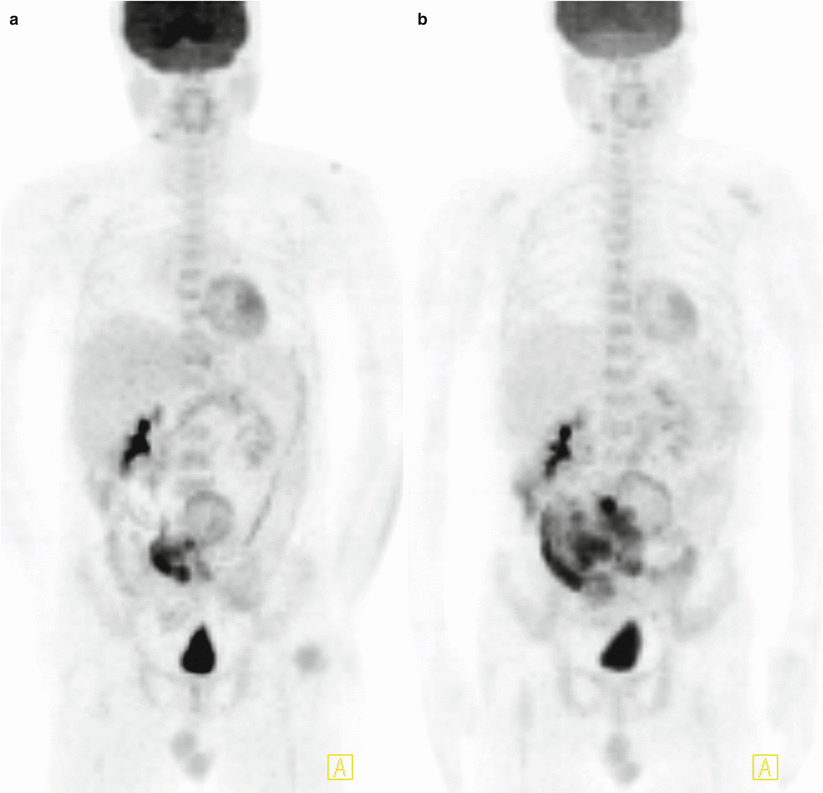
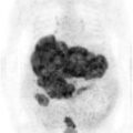
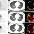
Fig. 12.1
MIP images show heterogenously hypermetabolic mass lesion located at lower abdomen (a), increase in size and metabolic activity of lesion 5 months after treatment (b)
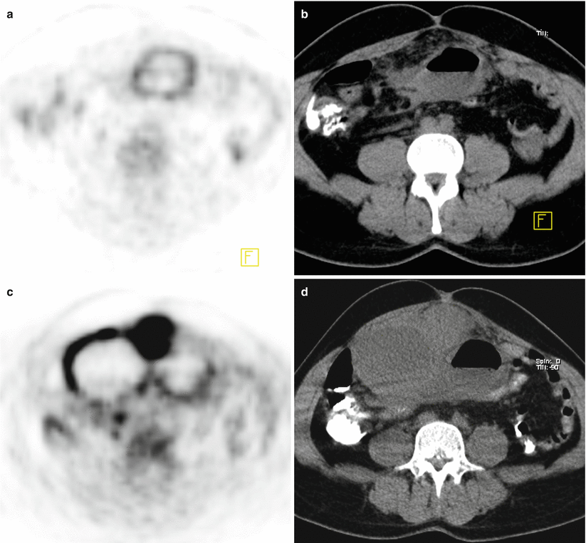
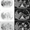
Fig. 12.2
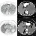
Axial PET and CT images show mass lesion with intensely increased FDG uptake in circular fashion, located adjacent to anterior wall of abdomen (SUVmax 13.5) (a, b). After Gleevec therapy the lesion increased in size with the inner side being more hypodense and intensity of FDG uptake increased at periphery of mass with a newly appearing solid component (SUVmax 14.7) (c, d)
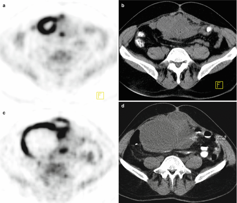
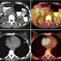
Fig. 12.3
Axial PET and CT images showing hypermetabolic mass lesion at the entrance of pelvis before (a) and after (b) Gleevec therapy (c, d)
Interpretation
The increase in size and metabolic activity of lesion is consistent with progression.
Teaching Point
18F-FDG PET/CT scan is effective in assessment of response to Gleevec therapy in GIST. Although regression is detected on PET, these tumors might become larger within the few months following the therapy, because of hemorrhage, edema, or myxoid degeneration. In this case the lesion became larger but since there is also increase in metabolic activity, the case is accepted as progression.
12.2 Case 2: Gastrointestinal Stromal Tumor Progression
History
A 57-year-old male who had the diagnosis of GIST after excision of a tumoral lesion from small intestine, underwent chemotherapy with Gleevec, and the efficacy of the therapy is being assessed with 18F-FDG PET/CT at regular intervals.
Findings
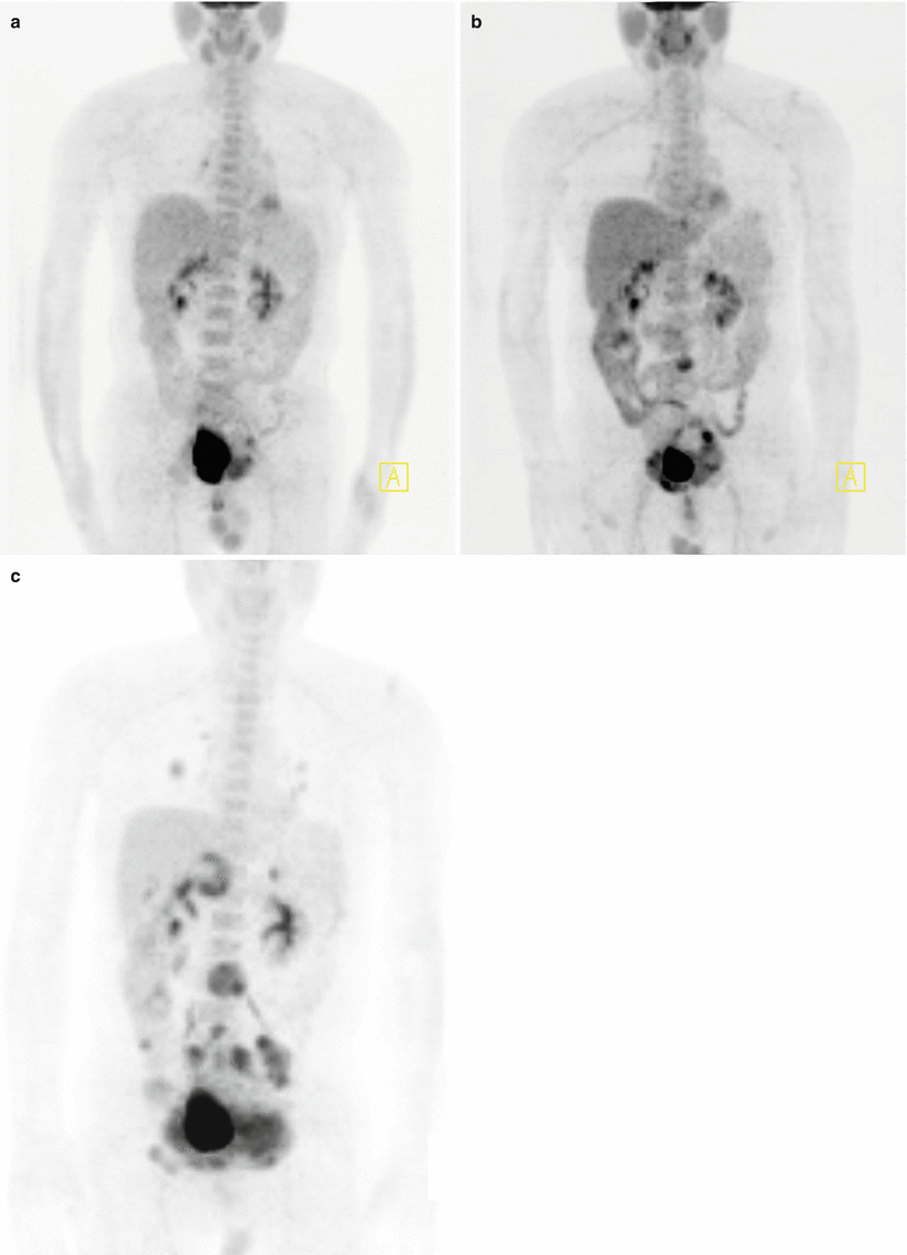
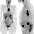
Fig. 12.4


MIP images show intensely increased FDG uptake at mass lesion in front of the bladder (SUVmax 7.2) (a), 10 months later slight regression at the lesion is seen (SUVmax 6.6) but there is a mildly hypermetabolic newly developed lesion at level of L3 (b), 7 months after the second study there is progression at the lesion (SUVmax 9.5) and newly developed numerous hypermetabolic lesions in abdomen and both lungs are noted (c)
Related posts:
Stay updated, free articles. Join our Telegram channel
Full access? Get Clinical Tree
