Pure form: Affects only pancreaticoduodenal groove
Segmental form: Affects pancreaticoduodenal groove and extends medially into pancreatic head
• Pancreatic groove is a theoretical space defined by pancreatic head (medially), 2nd portion of duodenum (laterally), 3rd portion of duodenum and IVC (posteriorly), and duodenal bulb (superiorly)
IMAGING
• Sheet-like, curvilinear soft tissue mass between pancreatic head and duodenum
May demonstrate delayed enhancement
MR: Usually T1 hypointense with variable T2 signal (depending on acuity)
• Thickened medial duodenal wall ± cysts within groove or thickened duodenal wall
• Segmental form: Mass-like enlargement of pancreatic head
• Calcifications and ductal dilatation/beading can develop in chronic setting (similar to traditional chronic pancreatitis)
TOP DIFFERENTIAL DIAGNOSES
• Pancreatic ductal adenocarcinoma
• Duodenal carcinoma
• Acute edematous pancreatitis involving groove
CLINICAL ISSUES
• Usually middle-aged men with history of alcohol abuse
• Amylase, lipase, and tumor markers are usually normal
• Prospective diagnosis is very uncommon; difficult to exclude malignancy with imaging or biopsy
• Surgery (Whipple procedure) may be required to rule out malignancy or due to intractable symptoms
(Left) Gross photo of a pancreaticoduodenectomy specimen shows a mass-like lesion beneath the duodenal mucosa, representing groove pancreatitis. Note the paraduodenal zone of fibrosis with numerous small cysts .
(Right) Axial CECT shows subtle soft tissue thickening in the pancreaticoduodenal groove, as well as mass-like pancreatic head enlargement with an internal cyst . This was found to be segmental (given pancreatic head involvement) groove pancreatitis at surgery.
(Left) Axial CECT image demonstrates a hypodense soft tissue mass in the pancreaticoduodenal groove with associated cystic spaces , resulting in upstream pancreatic ductal dilatation .
(Right) Coronal volume-rendered CECT image nicely demonstrates the hypodense soft tissue in the pancreaticoduodenal groove with associated cystic spaces . Initially suspected to represent malignancy, this was found to be groove pancreatitis at surgery.
TERMINOLOGY
Synonyms
• Cystic dystrophy of duodenal wall, pancreatic hamartoma of duodenal wall, periampullary duodenal wall cyst
Definitions
• Form of chronic pancreatitis affecting pancreaticoduodenal groove
Pure form: Affects only pancreaticoduodenal groove
Segmental form: Affects pancreaticoduodenal groove and extends medially into pancreatic head
• Pancreatic groove is a theoretical space defined by pancreatic head (medially), 2nd portion of duodenum (laterally), 3rd portion of duodenum and IVC (posteriorly), and duodenal bulb (superiorly)
Contains distal common bile duct (CBD), main/accessory pancreatic ducts, and major/minor papilla
IMAGING
General Features
• Best diagnostic clue
Curvilinear soft tissue between pancreas and duodenum
CT Findings
• Sheet-like, curvilinear soft tissue mass (with delayed enhancement) between pancreatic head and duodenum


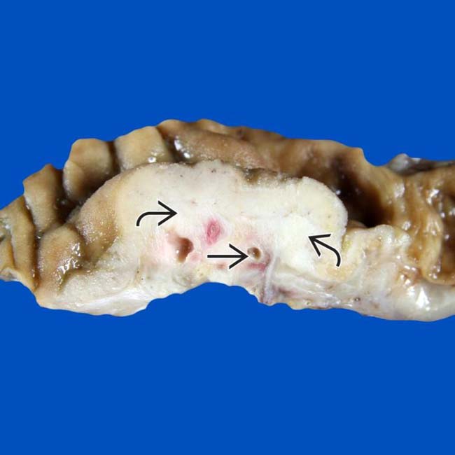
 beneath the duodenal mucosa, representing groove pancreatitis. Note the paraduodenal zone of fibrosis with numerous small cysts
  .
.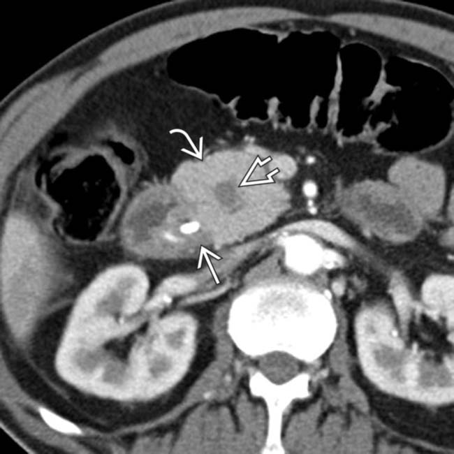
 in the pancreaticoduodenal groove, as well as mass-like pancreatic head
  enlargement with an internal cyst
  . This was found to be segmental (given pancreatic head involvement) groove pancreatitis at surgery.
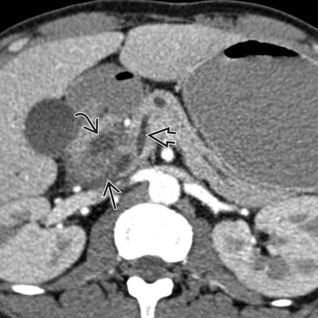
 in the pancreaticoduodenal groove with associated cystic spaces
  , resulting in upstream pancreatic ductal dilatation
  .
.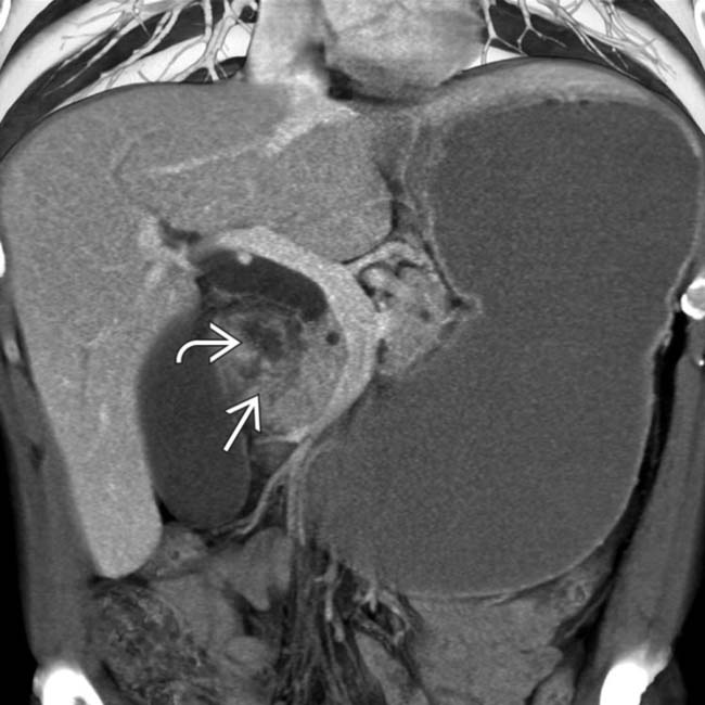
 in the pancreaticoduodenal groove with associated cystic spaces
  . Initially suspected to represent malignancy, this was found to be groove pancreatitis at surgery.


