Case presentation
A 15-year-old male presents to the emergency department with right wrist pain. The patient reports that he fell on his outstretched right hand while skateboarding. Physical examination is significant for mild swelling with tenderness to palpation over the distal right forearm. He has 2+ radial pulses with full range of motion and intact sensation in the radial, ulnar, and median distributions. Capillary refill in his fingers is normal. There is no shoulder or elbow pain, swelling, or deformity.
Imaging considerations
Plain radiography
Plain radiographs are the standard imaging modality for physeal fractures. Images should be obtained in at least two planes. Appropriate pain management is important in patients with potential fractures to help optimize positioning, making interpretation of the imaging study easier. There are cases in which a fracture may not be evident immediately on plain radiography. Nondisplaced Salter-Harris type I fractures will sometimes present with normal radiographs or have only subtle findings such as growth plate widening or surrounding soft-tissue swelling. Patients who continue to report tenderness over the growth plate on examination should be treated as if they have a Salter-Harris type I fracture with splinting of the extremity even in the setting of normal radiographs. These patients will require outpatient follow-up radiographs at 7 to 10 days, especially if they are still having pain, to assess for signs of bone healing response or other evidence of fracture that may not have been apparent on the original imaging studies. Salter-Harris type II–V fractures are usually apparent on plain radiographs.
Forearm physeal fractures in children can have an associated supracondylar fracture, so the lateral forearm view should include the distal humerus or there should be accompanying elbow views. If wrist imaging shows an isolated distal ulnar physeal fracture, further evaluation of the proximal radius should be obtained to rule out associated radial fracture or dislocation, as isolated ulnar physeal fractures are uncommon.
Computed tomography (CT)
Noncontrast CT imaging of physeal fractures is indicated only if needed for surgical planning or for further classification of Salter-Harris types III–V fractures depending on the site of injury. Discussion with an orthopedic specialist should occur before obtaining a CT in children with these fractures.
Ultrasound
Ultrasound is an emerging field for imaging pediatric fractures; however, plain radiographs continue to remain the gold standard for diagnosing physeal fractures. Ultrasound is currently indicated only as a first-line diagnostic modality in settings where plain radiography is not available. Ultrasound-guided forearm fracture reduction is also a rising trend, but more data are needed before this can be recommended over traditional reduction techniques.
Magnetic resonance imaging (MRI)
MRI can be useful in the outpatient setting to help differentiate acute injuries from chronic repetitive injuries in patients who have continued pain after expected healing, but it has limited diagnostic utility in the emergency department for suspected physeal injury.
Imaging findings
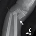
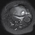
The patient had a two-view x-ray of his right forearm and a three-view x-ray of his right wrist. Selected images of the wrist series are provided. There is a displaced growth plate–related fracture of the distal right radius, appearing to be a Salter-Harris type II fracture; there is a nondisplaced oblique fracture of the proximal right first metacarpal; there is an ulnar styloid process fracture ( Figs. 51.1 and 51.2 ).


Case conclusion
The patient was treated with closed reduction under ketamine sedation after consultation with Pediatric Orthopedics, who performed the reduction procedure ( Figs. 51.3 and 51.4 ). The patient had a cast applied and follow-up scheduled with pediatric orthopedics seven days after his emergency department visit.


