and Filiz Özülker1
(1)
Nuclear Medicine, Okmeydani Training and Research Hospital, Istanbul, Turkey
3.1 Case 1: Adenoid Cystic Carcinoma of Lacrimal Gland
History
A 42-year-old female had undergone surgery for a mass in her left eye causing orbital pain and paresthesia and the pathology turned out to be adenoid cystic carcinoma of lacrimal gland. Patient was lost to follow up during 10 years period and eventually a 18F-FDG PET/CT study was performed.
Findings
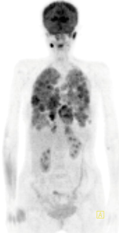
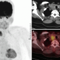
Fig. 3.1
MIP image shows intense and widespread FDG uptake at both lungs
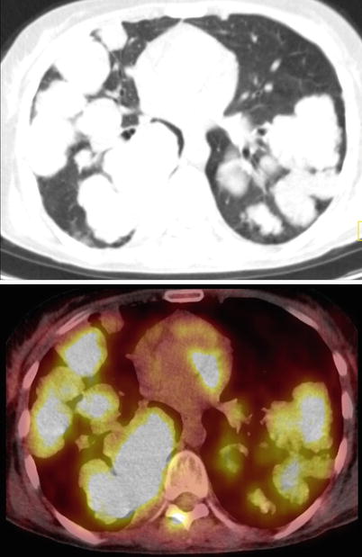
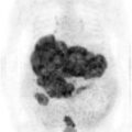
Fig. 3.2
Axial slices of CT and fusion images show mass lesions at both lungs with intense FDG uptake
Interpretation
Widespread metastatic lesions of adenoid cystic carcinoma at both lungs.
Teaching Point
Adenoid cystic carcinomas of the lacrimal gland are rare FDG avid malignant tumors. They are indolent tumors, which tend to spread to adjacent structures and occasionally metastasize via hematogenous spread to lungs, brain, and bone.
3.2 Case 2: Basal Cell Adenocarcinoma of the Right Parotid Gland
History
A 52-year-old male who was diagnosed with the basal cell adenocarcinoma of the right parotid gland underwent 18F-FDG PET/CT before and after surgical resection of the tumor.
Findings
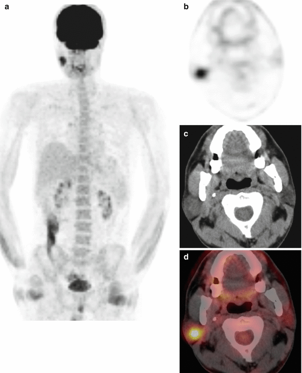
Fig. 3.3
MIP image (a), axial PET, CT, and fusion images show focal increased FDG uptake at right parotid gland (b–d)
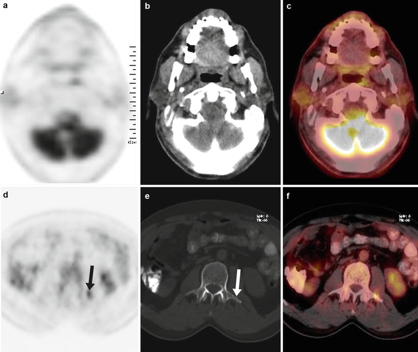
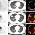
Fig. 3.4
18F-FDG PET/CT study performed while the patient is status post surgical resection of the lesion at right parotid gland shows the clearance of the pathological activity at right parotid gland (a–c), and FDG uptake at the left transverse process of Lomber 2 vertebra (arrow) corresponding to a fracture at this site (d–f)
Interpretation
18F-FDG PET/CT study showed the therapy response at right parotid gland tumor. The pathological uptake at the transverse process of Lomber 2 vertebra is due to simple fracture rather than being a metastasis.
Teaching Point
Basal Cell Adenocarcinoma of parotid gland is a rare disease entity and it is FDG avid.
Benign bone fractures should not be misinterpreted as metastasis while evaluating 18F-FDG PET/CT studies.
3.3 Case 3: Oncocytic Carcinoma of Parotid Gland
History
A 45-year-old female was diagnosed as oncocytic carcinoma of the right parotid gland and underwent 18F-FDG PET/CT for staging.
Findings
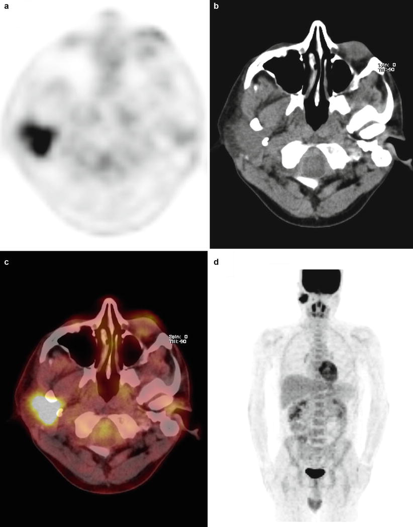
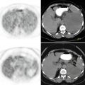
Fig. 3.5
Axial PET, CT, and fusion images (a–c) and MIP image (d) show intensely increased FDG uptake at right parotid gland
Interpretation
18F-FDG PET/CT study showed intensely increased FDG uptake at right parotid gland mass confirming the malignant nature of the lesion
Teaching Point
Oncocytic carcinoma is an extremely rare malignancy in salivary glands, accounting only for 0.5 % of all epithelial salivary gland malignancies. Oncocytic carcinoma is an FDG avid malignancy but one must bear in mind that benign pathologies of salivary glands like oncocytoma, pleomorphic adenoma, and Warthin tumor are also FDG avid.
3.4 Case 4: Therapy Response in Nasopharyngeal Carcinoma
History
A 34-year-old male with nasopharyngeal carcinoma was evaluated with PET/CT before and after having chemotherapy and radiotherapy.
Findings
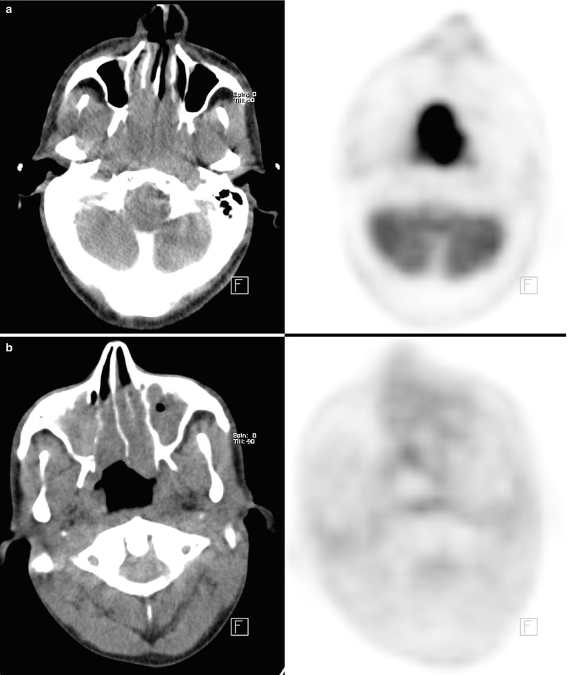
Fig. 3.6
Large mass with high FDG uptake (SUVmax 15) extending from right posterior nasal cavity to right posterolateral wall of nasopharynx (a). Axial slice of 18F- FDG PET/CT study performed 10 months after initial staging study shows clearance of the pathological uptake after treatment (b)
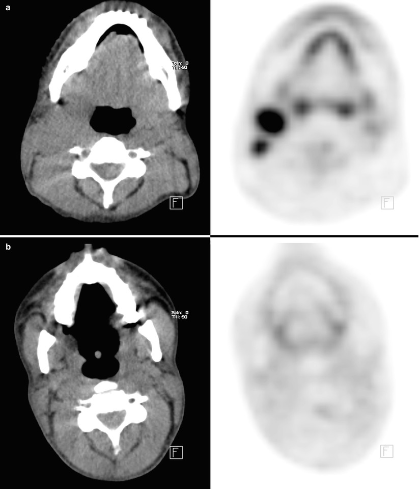
Fig. 3.7
Two intensely hypermetabolic foci (SUVmax 13) at the right level II lymph node station (a) Disappearance of the lesions after treatment (b)
Interpretation
Intensely hypermetabolic mass lesion at nasopharynx consistent with nasopharyngeal carcinoma and right cervical metastatic lymph nodes. All of the pathological activities are cleared after treatment indicating complete metabolic response to therapy.
Teaching Point
18F-FDG PET/CT can precisely assess response to therapy in nasopharyngeal carcinoma, which gives prognostic information for patients. The SUVmax corresponds well with response of tumor to treatment, with greater decreases in SUVmax correlating well with better survival.
3.5 Case 5: Detection of Recurrence in Tongue Carcinoma
History
A 55-year-old male underwent resection of tongue for tongue carcinoma and after surgery had a flap reconstruction of the tongue. The patient was referred for restaging purpose and a 18F-FDG PET/CT study was performed.
Findings
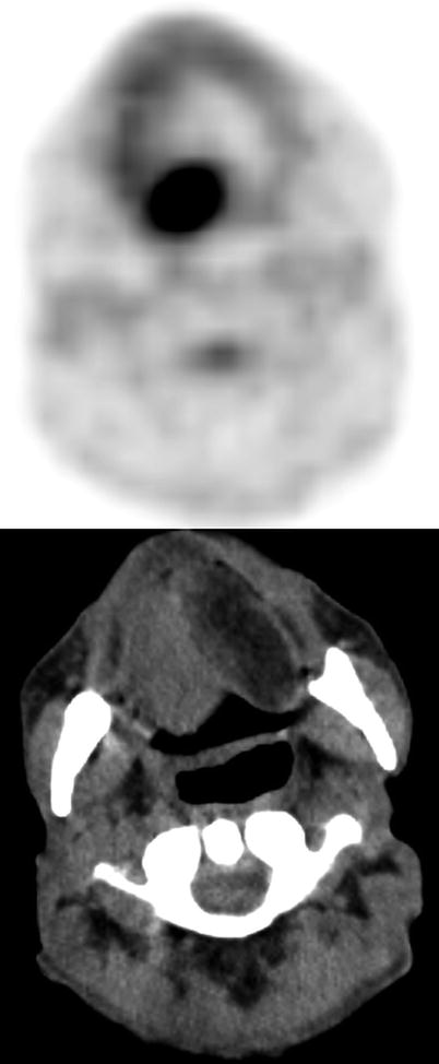
Fig. 3.8
Axial PET and CT images show intense focal FDG uptake at the posteromedial border of the flap
Interpretation
Extensive FDG uptake at the posteromedial border of the flap compatible with recurrence of the primary malignant disease.
Teaching Point
Recurrence of tumor may not be detectable clinically especially when the lesion is deep to the flap reconstruction. The thickening along the flap border due to postsurgical changes and granulation tissue may not be possible to be distinguished from recurrence with anatomical imaging methods. FDG PET/CT is effective in detecting the relapse of the tumor in such cases.
3.6 Case 6: Squamous Cell Carcinoma of the Epiglottis
History
A 75-year-old female had the diagnosis of squamous cell carcinoma of the epiglottis after a biopsy and underwent 18F-FDG PET/CT scan for pretreatment staging.
Findings
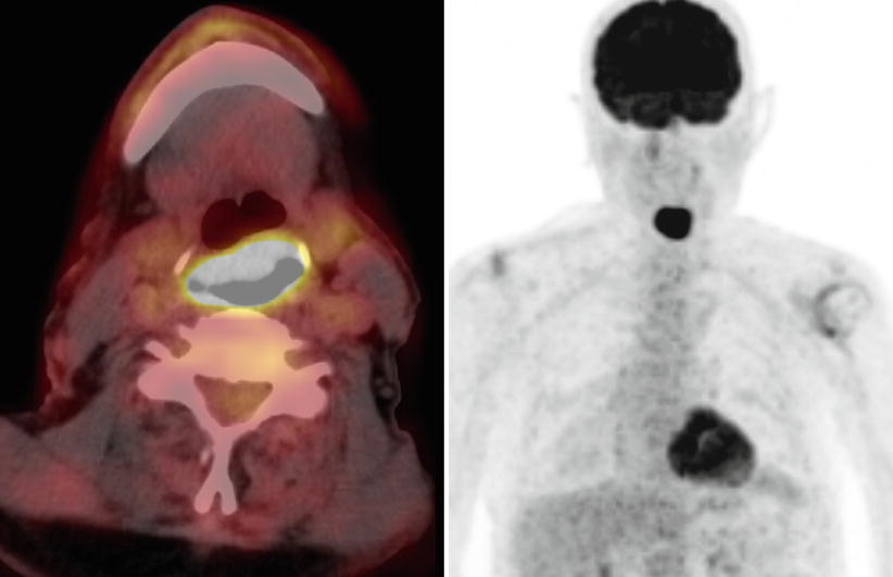
Fig. 3.9
Axial fusion and MIP images show intensely hypermetabolic lesion (SUVmax 12.9) at epiglottis. There is not any detectable cervical lymph node showing pathologically increased FDG uptake
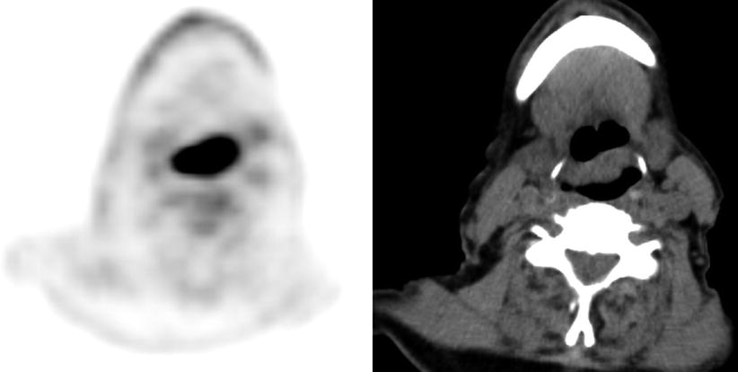
Fig. 3.10


Axial PET and CT images show intensely increased FDG uptake at epiglottis
Related posts:
Stay updated, free articles. Join our Telegram channel
Full access? Get Clinical Tree
