38.1 Axial CT brain: normal
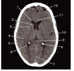
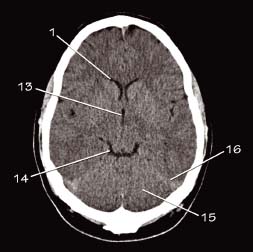
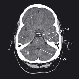
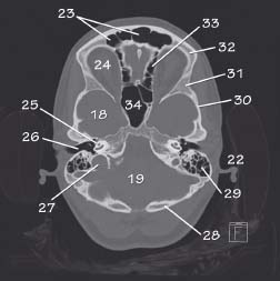
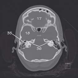
Key
| 1 Lateral ventricle (anterior horn) |
| 2 Caudate nucleus |
| 3 Internal capsule |
| 4 White matter |
| 5 Grey matter |
| 6 Lateral ventricle (posterior horn) |
| 7 Falx cerebri |
| 8 Choroid plexus (calcified)temporal bone |
| 9 Corpus callosum (splenium) |
| 10 Sylvian fissure |
| 11 Corpus callosum (genu) |
| 12 Sulci |
| 13 Third ventricle |
| 14 Basal cisterns |
| 15 Cerebellum |
| 16 Tentorium cerebelli |
| 17 Anterior cranial fossa |
| 18 Middle cranial fossa |
| 19 Posterior cranial fossa |
| 20 Fourth ventricle |
| 21 Pons |
| 22 Pinna of ear |
| 23 Frontal air sinuses |
| 24 Orbit |
| 25 Cochlea within petrous |
| 26 External auditory meatus |
| 27 Jugular foramen |
| 28 Occipital bone |
| 29 Mastoid air cells |
| 30 Squamous temporal bone |
| 31 Greater wing of the sphenoid |
| 32 Frontal bone |
| 33 Ethmoid air cells |
| 34 Sphenoid sinus |
| 35 Semicircular canal |
| 36 Internal auditory meatus |
Related posts:


Stay updated, free articles. Join our Telegram channel

Full access? Get Clinical Tree


