Only 50% of hepatic angiomyolipomas have substantial fat component
• Arterial phase: Prominent enhancement of nonfatty portion of lesion
Central vessels within lesion if mass is large
• Fatty component of tumor results in hyperintense (high signal) foci on T1WI and T2WI
• MR, fat suppression ± opposed-phase GRE imaging
TOP DIFFERENTIAL DIAGNOSES
• Hepatocellular carcinoma
• Postoperative state, liver
• Focal steatosis
• Hepatic adenoma
• Hepatic lipoma
• Metastases
Teratoma or liposarcoma
PATHOLOGY
• Associated with tuberous sclerosis in < 10% of cases
But some patients likely have forme fruste tuberous sclerosis
DIAGNOSTIC CHECKLIST
• Small, fat density hepatic mass in patient with tuberous sclerosis is almost certainly benign
• Angiomyolipoma that is primarily myeloid or angioid may be indistinguishable from other hepatic tumors, including hepatocellular carcinoma
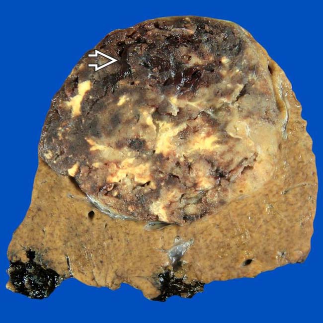
(Left) Gross photograph of a fixed specimen shows a heterogeneous mottled tan, yellow, and brown tumor with areas of hemorrhage and degeneration . Note that the background liver is not cirrhotic. (Courtesy J. Misdraji, MD.)
(Right) H&E-stained section shows a tumor composed of 3 elements: Adipose tissue , vessels , and plump spindle cells . (Courtesy J. Misdraji, MD.)
(Left) Axial T2WI MR shows a heterogeneously bright mass with a fatty component that is nearly isointense to subcutaneous fat . The rest of the tumor has the moderate hyperintensity typical of most neoplasms on T2WI MR.
(Right) Axial T1WI MR in the same patient shows that most of the mass is hyperintense, an unusual feature of most neoplasms and generally indicative of the presence of fat or hemorrhage within the mass. This tumor was resected and proved to be an isolated angiomyolipoma (AML).
TERMINOLOGY
Abbreviations
• Hepatic angiomyolipoma (AML)
Synonyms
• Benign hepatic hamartoma
Definitions
• Benign mesenchymal tumor composed of variable amounts of smooth muscle (myoid), fat (lipoid), and proliferating blood vessel (angioid) components
IMAGING
General Features
• Best diagnostic clue
Well-circumscribed, mostly fatty mass in liver
• Location
Liver is 2nd most common site (kidney is 1st)
• Size
Variable; 0.3-36 cm in diameter
• Key concepts
Round or lobulated solitary mass or multiple lesions with variable shape
Only 50% of hepatic angiomyolipomas have substantial fat component
– Those without much fat are difficult to distinguish from other hepatic tumors
CT Findings
• NECT
Well-defined mass with heterogeneous attenuation values due to presence of fat and soft tissue densities
– May be almost completely fat or soft tissue density mass
• CECT
Arterial phase: Prominent enhancement of nonfatty portion of lesion
Portal phase: Lesion shows hypoattenuation throughout mass
• CTA
Central vessels within lesion if mass is large
MR Findings
• T1WI
Hypointensity or hyperintensity on T1WI
– Depends on amount of fat and whether fat-suppressed technique is used
Only gold members can continue reading. Log In or Register to continue


 . Note that the background liver is not cirrhotic. (Courtesy J. Misdraji, MD.)
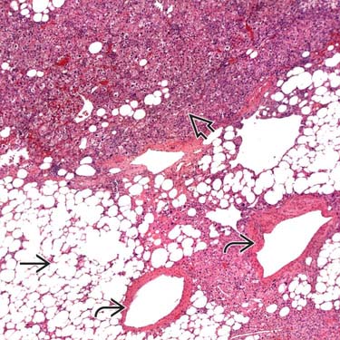
 , vessels
  , and plump spindle cells
  . (Courtesy J. Misdraji, MD.)
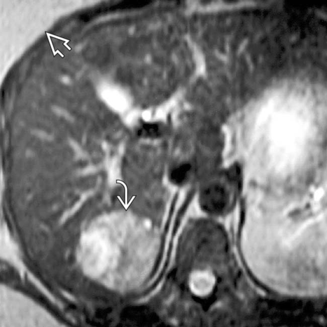
 that is nearly isointense to subcutaneous fat
  . The rest of the tumor has the moderate hyperintensity typical of most neoplasms on T2WI MR.
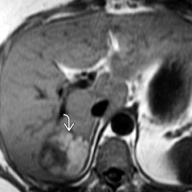
 is hyperintense, an unusual feature of most neoplasms and generally indicative of the presence of fat or hemorrhage within the mass. This tumor was resected and proved to be an isolated angiomyolipoma (AML).


 Hypointensity or hyperintensity on T1WI


