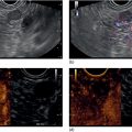
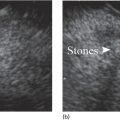
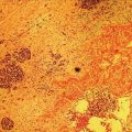
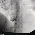
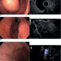
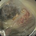
Spencer Cheng, Mauricio K. Minata, Carlos K. Furuya, and Edson Ide Department of Gastroenterology, Gastrointestinal Endoscopy Unit, University of São Paulo, São Paulo, Brazil Learning endoscopic ultrasound (EUS) can be challenging since it is one of the most difficult endoscopic techniques. Generally, a long learning curve is necessary to master EUS. A variety of strategies have been developed to improve mastery of EUS, respecting the principle of achieving a high level of expertise without causing harm to the patient. There is a necessity to develop experimental models to teach EUS considering the great demand for learning, training and testing new techniques and accessories. Currently available models for EUS training include computer‐based simulators, tabletop phantom models, ex vivo models, and live animal models. In this chapter, we cover topics concerning ex vivo models. The EASIE model was the first and most widely used as the basis for all current ex vivo models employed for training of endoscopic therapeutic techniques. Later, the EUS RK model was developed, based on improvements in the EASIE model by adding miscellaneous substance molds to simulate extraparietal lesions. This model has become a reference for training EUS puncture techniques of subepithelial, hepatic and lymph node lesions, and drainage of collections with self‐expanding metal or plastic stents. Further progress was made when the use of animal models showed a good correlation with human anatomy for EUS. Artifon et al. adapted porcine ex vivo models for training EUS puncture with good acceptance and reproducibility. The study of anatomy with EUS and the diagnosis and puncture of lesions can be performed using the extracted viscera consisting of esophagus, stomach, duodenum, jejunum, liver, and gallbladder, fixed in a box (Figure 52.1). Vascular structures can be simulated using a segment of small bowel filled with water and positioned near the stomach of ex vivo models (Figure 52.2). Very simple materials, such as chicken tenderloin, grapes, spleen, omentum fragments, and gel, can be used to create models. These models can simulate lymph nodes, hepatic nodules and subepithelial lesions (Figures 52.3 and 52.4). Some other simple, fast, inexpensive and easily reproduced experimental models have been described for simulation of pancreatic pseudocysts and fluid collections. Cysts can be simulated with a porcine bladder filled with ultrasound gel and the model used to practice EUS fine needle aspiration (FNA) and pseudocyst drainage (Figures 52.5 and 52.6
52
How to use ex vivo Models in Teaching Therapeutic Endoscopic Ultrasound
Introduction
Ex vivo models
![]()
Stay updated, free articles. Join our Telegram channel

Full access? Get Clinical Tree


