Key Facts
- •
Hypophosphatasia is an autosomal recessive disorder.
- •
Radiographic findings of osteomalacia or rickets are present without abnormalities of vitamin D metabolism.
- •
High levels of phosphoethanolamine are noted in the blood and urine.
- •
In the neonatal form, transverse bony spurs (Bowdler spurs) may be present under skin dimpling.
- •
Craniosynostosis may result in a small skull.
- •
In contrast to most causes of osteomalacia in which Looser zones appear in the inner cortex, hypophosphatasia presents with Looser zones in the outer cortex of long bones.
Hypophosphatasia, an autosomal recessive disorder, is one of the rare syndromes in which the radiographic findings of osteomalacia or rickets are present in the absence of abnormalities of vitamin D metabolism. Serum levels of calcium and phosphate are high or normal. The serum alkaline phosphatase is low and there are abnormal amounts of phosphoethanolamine in the urine and blood.
MUSCULOSKELETAL FINDINGS
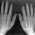
The severity of radiologic findings depends on the age of presentation and increases as the disease presents in younger ages. Radiographs show generalized osteopenia with a coarse trabecular pattern, healing or non-healing fractures, bowing deformities (especially involving the tibiae), chondrocalcinosis, craniosynostosis (resulting in a small skull), early loss of deciduous teeth, and subperiosteal bone formation. In the neonatal form, transverse bony spurs from the radius, ulna, and fibula (Bowdler spurs) may be present under skin dimpling.
Severe osteopenia is observed in the calvarium; however, the appearance of the skull base is more normal. Cranial sutures may appear widened due to undermineralization.
Growth plate changes resemble those seen in rickets with characteristic irregular and prominent tongue-like radiolucent extensions into the metaphyses of the distal femurs, proximal humeri, epiphyses, and carpal bones.
Fractures occur following minor trauma, and minimal callus may be seen with delayed healing. In contrast to most causes of pure vitamin D-deficient osteomalacia in which Looser zones involve the inner cortex, hypophosphatasia presents with Looser zones in the outer cortex of long bones. Multiple rib fractures and abnormal appearance of the distal phalanges are other radiographic manifestations.
Tendinous and ligamentous insertions in the paravertebral regions may show ossification.
Related posts:


Stay updated, free articles. Join our Telegram channel

Full access? Get Clinical Tree


