Key Facts
- •
The three-phase bone scan consists of (1) an early blood flow phase that reflects vascularity, (2) a blood pool phase that shows the level of soft tissue involvement, and (3) delayed images (2 to 4 hours after injection) that reflect the osteoblastic response to the underlying disease.
- •
A normal bone scan essentially excludes osteomyelitis.
- •
Indium white blood cell scanning requires in vitro labeling of 40 mL of the patient’s blood for imaging.
- •
Gallium scans are interpreted by comparison to bone scan images; if gallium uptake exceeds that of the bone scan uptake or differs in distribution from the area of bone scan uptake (termed incongruent uptake ), then osteomyelitis is likely.
- •
Unlike the bone scan, Gallum-67 citrate activity returns to baseline approximately 6 weeks after successful treatment of osteomyelitis and can therefore be used to monitor the clinical course of the disease.
- •
Indium scanning may be falsely negative in patients with vertebral osteomyelitis and discitis.
- •
Magnetic resonance imaging (MRI) is more sensitive and specific than scintigraphy for identifying radiographically occult scaphoid fractures.
- •
In elderly patients, it may take several days for an acute fracture to be seen on a bone scan.
- •
After a fracture, the delayed images of a bone scan may remain positive for years; an abnormal scan has been reported as long as 40 years.
- •
A healing “flare phenomenon” has been described as increased radiotracer uptake in an area of previously noted skeletal metastasis on a bone scan associated with increased sclerosis on radiographs or CT scan. A flare phenomenon is usually seen during the first 3 months after chemotherapy and represents a favourable response to therapy.
PRINCIPLES OF MUSCULOSKELETAL SCINTIGRAPHY
Nuclear medicine plays an important role in the diagnosis and management of various skeletal diseases. Bone scanning reflects changes in bone physiology in response to underlying disease. Diphosphonate compounds (methylene diphosphonate [MDP], hydroxymethylene diphosphonate [HDP]) labeled with technetium-99 (Tc-99m) are the radiotracers of choice for routine bone scintigraphy. In addition to diphosphonates, other radiopharmaceuticals are useful for skeletal imaging ( Table 2-1 ).
| Radiotracer | Physiologic Half-Life | Mode of Photon Decay | Energy (kev) | Mode of Production | Critical Organ |
|---|---|---|---|---|---|
| Tc-99m diphosphonate MDP/HDP | 6.0 hours | IT | 140 | Reactor | Bladder |
| In-111 oxine | 2.81 days | EC | 172, 247 | Accelerator | Spleen |
| Gallium-67 citrate | 78.1 hours | EC | 93,185, 300, 394 | Accelerator | Large bowel |
| F-18 FDG | 110 minutes | B+ | 511 | Accelerator | Bladder |
| Thallium 201 chloride | 73.1 hours | EC | 81 | Accelerator | Renal cortex |
Technical Considerations
Radiopharmaceuticals such as Tc-99m MDP or Tc-99m HDP should be used within 2 hours and no later than 6 hours after preparation. These compounds decompose with time due to the oxidation-reduction process and result in excess free pertechnetate that may degrade the image. The recommended dose of Tc-99m MDP is 20 to 25 millicurie (mCi) (750 to 900 megabecquerel [MBQ]). Hydration of the patient before imaging is useful; it is suggested that the patient drink 4 to 6 glasses of water between injection of the isotope and imaging. The time of imaging depends on age; in patients younger than 20 years, imaging is done 2 hours after injection, and in older patients, a 3- to 4-hour delay is recommended to provide better image quality.
Imaging Technique
Whole body anterior and posterior images are routinely performed. These may be supplemented by single photon emission computed tomography (SPECT) images or pinhole (high resolution) views of a hip, wrist, or ankle for further evaluation of an area of interest. Sedation for patients younger than 4 years or older patients who are mentally challenged is recommended. When scans are interpreted, consideration is given to the positioning of the patient under the camera, the clinical information, and the biodistribution of the radiotracer; lack of knowledge about any of these factors may lead to false-positive or false-negative readings.
What Is SPECT?
SPECT (SPET in Europe) is a routine technique used in nearly any nuclear medicine department. With SPECT, the gamma camera (either single- or multi-head camera) moves around, viewing the patient from at least 180 degrees. For musculoskeletal imaging, 360 degrees of rotation is required for accurate image reconstruction; 180 degrees of rotation is usually used for cardiac imaging. The data set after SPECT imaging is reconstructed by filtered-back projection methods. The SPECT slices are viewed in the transverse, sagittal, or coronal planes or as three-dimensional (3D) representation of the organ. The main advantage of SPECT is the ability to view the reconstructed image in multiple planes and to separate overlapping structures. As much as a sixfold increase in image contrast can be obtained with SPECT. The anatomic location of various areas of increased or decreased radiopharmaceutical uptake can be better defined spatially. Recent technologic advances of SPECT imaging provide fusion of SPECT images with computed tomography (CT) or magnetic resonance imaging (MRI) to better identify the location of lesions.
What Is a Pinhole Collimator?
The selection of a particular type of collimator is made primarily on the basis of the size or area of the organ to be imaged and on the degree of detail desired in the anatomy. When the target area is not too large and higher-resolution scintigraphy and greater detail are desired, the pinhole collimator is used. A pinhole collimator is a cone-shaped lead shield, which tapers into a small aperture perforated at the center of the tip at a distance from the detector face. The geometry of the pinhole creates an inverted image of the target organ in the detector from the photons traveling through a small aperture. Any change in pinhole collimator design can affect lesion detectability by altering the spatial resolution and sensitivity. Common indications for pinhole imaging of the skeleton are the evaluation of Legg-Perthes disease (osteonecrosis of the hip in children) and better localization of fractures of the small bones in the wrist or ankle. In general nuclear medicine, the pinhole collimator is used routinely for imaging the thyroid or for renal cortical scintigraphy using technetium (dimercaptosuccinic acid) (Tc-DMSA) in cases of pyelonephritis.
What Is a Three-Phase Bone Scan?
The three-phase bone scan is primarily used to differentiate cellulitis from osteomyelitis. It can also be used for the diagnosis of osteoid osteoma, acute stress fracture, and reflex sympathetic dystrophy (RSD) ( Box 2-1 ).
Cellulitis versus osteomyelitis
Suspected osteoid osteoma
Suspected acute stress fracture
Suspected reflex sympathetic dystrophy
Acquisition
Images are obtained at specific time periods after isotope injection to demonstrate different physiologic information. Three phases are usually described ( Table 2-2 ).
Phase one: A dynamic flow study (radionuclide angiogram) consists of images obtained at 1-second intervals for 60 seconds after the intravenous injection of the radiopharmaceutical. The egion of interest should be within the camera’s field of view.
Phase two: Immediately following the angiogram, static (“blood pool”) images are obtained of the specific area or, when the localization of a lesion is not clear, a whole body image can be acquired.
Phase three: Delayed images are acquired 2 to 4 hours after injection of radiopharmaceutical.
| Phase | Name | Time after Injection | Purpose |
|---|---|---|---|
| One | Radionuclide angiogram (Blood flow) | 2 to 4 hours | Reflects vascularity |
| Two | Blood pool | Few minutes | Reflects soft tissue involvement |
| Three | Delayed | Immediate | Reflects osteoblastic response |
Interpretation
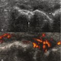
Blood flow images reflect the vascular supply (vascularity), blood pool images show the level of soft tissue involvement, and delayed images indicate osteoblastic response to the underlying disease ( Figure 2-1, A to C ).


What Is PET Imaging?
Positron emission tomography (PET) is a functional imaging technique using radionuclides (frequently F-18 FDG [fluoro-2-deoxy-D-glucose]) that measure the metabolic activity of the cell. This technique has broad clinical applications in oncology as well as for evaluation of neurologic and cardiac disorders. Oncology patients are asked to fast 4 to 6 hours prior to the examination.
A recent advance that combines functional imaging and CT, the PET/CT scanner, has reduced the study duration by eliminating the lengthy PET transmission scan and also provides accurate anatomic localization of functional abnormalities. A PET scanner provides resolution in the 7-mm to 10-mm range. Because the urinary bladder wall is the critical organ, hydration and frequent emptying of the bladder are recommended practices.
There are no absolute contraindications for PET imaging. A relative contraindication is pregnancy. Breastfeeding can resume 24 hours after isotope injection.
SCINTIGRAPHY OF INFLAMMATORY BONE DISEASES
Rheumatoid Arthritis
Accurate detection of the early synovitis in rheumatoid arthritis (RA) and other destructive inflammatory joint diseases is important to establish the most appropriate treatment and indicate prognosis. There has been no gold-standard study for the detection of synovitis activity ; MRI may be developing into a gold standard, but it is limited in the number of joints that can be assessed at one time. Bone scintigraphy is sensitive but not specific for the diagnosis of RA. In the acute stage of RA, the three-phase bone scan is positive. In the subacute and chronic stages of the disease, there is symmetric increased uptake, especially on delayed images, around affected joints ( Figure 2-2 ).

Labeled leukocytes are a promising method to evaluate the activity of the disease. The applicability of Tc-99m hexamethylpropylene amine oxime (HMPAO) labeled leukocyte scintigraphy in assessment of disease activity was tested in 21 patients with RA by Goal et al. In this study, significant correlation was found between labeled leukocyte accumulation in the hands and feet and clinical assessment of joint activity. F-18 FDG PET as a metabolic imaging device might be able to detect early inflammation. In addition, PET allows quantitation of F-18 FDG uptake, by which means disease activity can be assessed.
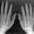
Osteoarthritis
Osteoarthritic changes can usually be demonstrated on standard radiographs. The blood flow and blood pool images of the bone scan usually do not show significant uptake. Delayed images, however, show increased periarticular uptake in a distribution typical of osteoarthritis (e.g., the thumb bases). Such findings are often incidentally seen on bone scans. The degree of uptake is proportional to the severity of disease.
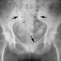
Sacroiliitis
The role of scintigraphy in the evaluation of patients with sacroiliitis is controversial. Planar and SPECT bone scintigraphy and Tc-99m sulfur colloid scanning with quantitative methods have all been used for the diagnosis. A combination of bone (Tc-99m MDP) and bone marrow (Tc-99m sulfur colloid) scanning may be useful in patients with active sacroiliitis. Increased Tc-99m MDP uptake with decreased or normal sulfur colloid uptake was the most common scintigraphic pattern in the acute phase of sacroiliitis in one study in which radiographic findings were normal or only slightly abnormal. SPECT bone scan was found to have the best accuracy (97% sensitivity, 90% specificity).
SCINTIGRAPHY IN SKELETAL INFECTION
When confronted with the potential diagnosis of an early skeletal infection, both morphologic and functional imaging modalities are frequently employed. The choice of imaging modality often depends mainly on whether or not the bone has been previously affected by another disease or surgery.
Ga67-Citrate Imaging
Unlike the bone scan, Ga-67 citrate activity returns to baseline approximately 6 weeks after successful treatment of osteomyelitis and can therefore be used to monitor the clinical course of the disease. The sensitivity of Ga-67 for acute osteomyelitis is 80% to 85%. Positive gallium scans may also be seen with primary skeletal tumor, skeletal metastasis, chronic infection, and septic inflammatory and traumatic lesions ; therefore specificity is low, approximately 70%. To improve the specificity, Tumeh et al. suggested that osteomyelitis is more likely to be present when gallium uptake exceeds that of Tc-99m MDP (bone scan) uptake or differs in distribution (termed incongruent uptake) ( Figure 2-3, A to C ). If gallium uptake is less than that of Tc-99m MDP uptake, infection is unlikely. If uptake on both exams is equivalent, the finding may be indeterminant, a circumstance seen mainly with preexisting abnormalities such as diabetes or posttraumatic changes. Gallium scanning may not be able to differentiate osteomyelitis from neuropathy or healing fracture ; in these conditions, In-111 white blood cell (WBC) scanning is useful. SPECT gallium and SPECT bone scans are more sensitive than planar gallium and planar bone scans specifically for vertebral osteomyelitis.


In-111 Leukocyte Imaging
In-111 oxine and Tc-99m HMPAO labeled leukocyte imaging are widely used in the diagnosis of osteomyelitis. An overall sensitivity of 88% and specificity of 91% are reported for osteomyelitis. These studies are especially useful in excluding infection in a previously violated bone such as may occur from prior trauma, postsurgery, or with diabetes ( Figure 2-4, A to D ). False-positive scans have been reported with recent trauma or acute fracture or following arthroplasty, and the addition of bone marrow scanning may be needed in these situations. Delay in the injection of the labeled leukocytes may cause false-negative results mainly due to decreased leukocyte viability when out of the body for a prolonged time.


Bone scanning should be performed in conjunction with labeled leukocyte imaging for anatomic localization. Because a negative bone scan essentially rules out osteomyelitis, there is no need to proceed with WBC scan if the bone scan is normal. Tc-99m HMPAO labeled leukocyte scanning has been reported to yield an accuracy similar to that of In-111 WBC scanning in the diagnosis of osteomyelitis but has the additional benefit of providing same-day results. This technique may be particularly useful in children, as the radiation dose is much lower than that of the In-111 leukocyte technique. A disadvantage of the Tc-99m HMPAO scan is the inability to acquire dual Tc-99m MDP and Tc-HMPAO leukocyte scans simultaneously.
A negative bone scan essentially rules out osteomyelitis; there is no need to proceed with a WBC scan if the bone scan is normal.
In-111 leukocyte scanning is not generally useful in the diagnosis of vertebral osteomyelitis; the images may show normal or decreased uptake even when osteomyelitis is present. This may be related to the chronic nature of the disease due to delayed diagnosis. Gallium scanning is the modality of choice for vertebral osteomyelitis and discitis.
Tc-99m Bone Marrow Scintigraphy
The addition of Tc-99m sulfur colloid scanning to the WBC scan protocol improves specificity for infection in complicated cases such as postarthroplasty infections. Infection is confirmed when there is less or no bone marrow activity on the sulfur colloid scan in areas with increased uptake on the labeled WBC scan. Activity present on bone marrow scans equal to or greater than that of the WBC scan indicates physiologic bone marrow activity and rules out infection ( Figure 2-5, A to C ).

Infection is confirmed when there is no activity or less bone marrow activity on the sulfur colloid scan in areas in which there is increased uptake on the labeled WBC scan.
Tc-99m Diphosphonate Imaging (Tc-99m MDP or HDP)
Three-phase bone scanning is the imaging modality of choice for suspected osteomyelitis. This examination becomes positive within 24 hours after onset of symptoms. The classic findings of osteomyelitis on three-phase bone scan are increased regional perfusion as seen in blood flow (phase 1) and blood pool (phase 2) and corresponding focal increased uptake on delayed images (see Figure 2-1 ). Pinhole imaging can be of value in children for better characterization of delayed focal uptake or for the small bones in adults, such as the wrist and ankle. This uptake pattern is different from cellulitis, which shows regional or diffusely increased uptake in the area involved on phase 1 and phase 2 but either no corresponding uptake on delayed images or only mildly increased uptake secondary to the hyperemia of adjacent or surrounding soft tissue infection.
Cellulitis shows regional or diffusely increased uptake in the area involved on phase 1 and phase 2 with either no corresponding uptake on delayed images or only mildly increased uptake secondary to the hyperemia. Osteomyelitis shows increased uptake in all three phases of the bone scan.
Bone scanning is very specific for the early diagnosis of osteomyelitis when the bone is not previously affected by other pathologic conditions (nonviolated) and is an efficient and cost-effective modality in the diagnosis. Overall sensitivity and specificity of bone scan for osteomyelitis in nonviolated bone are 90% and 95%, respectively. However, there have been some reports of false-negative scans in cases with proven early acute osteomyelitis, demonstrating either reduced or normal accumulation of the radiotracer, particularly in neonates. Cold lesions on bone scan may indicate a more virulent infectious process and are thought to be secondary to increased intraosseous and subperiosteal pressure, likely due to edema. Tuson et al. found that the positive predictive value of reduced uptake (cold lesion) in a selected group of patients was higher (100%) than that of a hot lesion (82%). Gallium scan or indium WBC scan may be helpful. A normal-appearing bone scan at a region of osteomyelitis may be due to its being obtained during the transition from cold to hot phases; otherwise, a negative three-phase bone scan can rule out osteomyelitis, and no further imaging is required. In violated bone, the bone scan alone may not establish the diagnosis, requiring complementary radionuclide imaging such as gallium or In-111 WBC scanning to improve the specificity.
F-18 FDG PET
F-18 FDG PET plays a major role in the field of oncology; however, its utility in infection and inflammatory lesions has also been described. Love et al. compared F-18 FDG with WBC/bone marrow scan (BMS) imaging. PET was 100% sensitive (94% for WBC/BMS) but its specificity was very low at 11% (100% for WBC/BMS). Further studies are needed to evaluate the efficiency of PET imaging in osteomyelitis. Thus far, the In-111 WBC/BMS combination remains the gold standard for the diagnosis of osteomyelitis.
Scintigraphy of Infected Prostheses
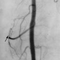
The distinction between mechanical failure of a prosthesis and infection may be difficult clinically, radiographically, and on scintigraphy. There is no specific scintigraphic pattern that indicates infection versus loosening. Furthermore, the pattern of normal postoperative increased uptake that occurs due to bone remodeling varies considerably depending on the type of prosthesis used and the time since surgery.
In the case of total hip arthroplasty (THA), knowledge of the type of implant and implant fixation are important to plan a diagnostic strategy. In cemented THAs, most asymptomatic patients show no significant periprosthetic uptake 6 months or more after operation. Focal uptake at the tip of a femoral component after 6 months indicates loosening. In a symptomatic patient, diffuse uptake around the shaft is thought to be more in favor of infection, and further evaluation with In-111 WBC is needed. In noncemented THA, postoperative periprosthetic uptake may continue for 2 years or longer in asymptomatic patients ( Figure 2-6, A, B ). In these noncemented THAs, focal uptake at the tip of a femoral component may indicate remodeling due to mechanical changes in the adjacent bone rather than loosening (see Figure 2-6 ).