Key Facts
- •
Characteristics of fatigue fractures include: (1) the activity is new or different for the individual; (2) it is strenuous; and (3) the activity is repeated with a frequency that ultimately produces symptoms.
- •
Insufficiency fractures result from normal or physiologic muscular activity on a bone that is deficient in mineral or in elastic resistance.
Stress injuries of bone comprise a constellation of abnormalities that have as their common denominator overuse of the involved bones. This category of diseases includes stress fractures, the most common of the group; toddler’s fractures; and “shin splints.” Classically, stress fractures may be divided into two broad categories: fatigue fractures and insufficiency fractures. Fatigue fractures are the result of overuse of bones of normal mineralization; insufficiency fractures result from normal activity on bones that are either deficient in mineral (the most common) or have abnormal mineralization (e.g., Paget’s disease).
Fatigue fractures result from overuse on bones of normal mineralization; insufficiency fractures result from normal activity on bones that are either deficient in mineral or have abnormal mineralization.
Toddler’s fractures are a variant of stress fractures because they occur in infants learning how to walk, at an age when their bones are not sufficiently mineralized to take the loading that is placed upon them. Shin splints is a generic term for any kind of pretibial pain that may result from overactivity. The actual injuries suffered by patients so afflicted may range from muscle or fascial tears to ligament strain, or stress fracture.
Stress injuries are common and are specific in both their location, as well as in regard to the activity that produced them. Although stress injuries are common, the diagnosis is often delayed because trauma is frequently not considered as the etiology for these lesions. The wide interest in physical fitness over the past 2 decades, however, has resulted in greater attention being paid to stress injuries. Furthermore, insufficiency fractures are receiving greater attention, particularly as they frequently occur in patients with known malignancy.
This chapter describes the pertinent aspects of stress injury to bone, focusing on those entities commonly encountered and the role that diagnostic imaging plays in establishing the diagnosis. Particular attention will be placed upon the use of magnetic resonance imaging (MRI), which has not only a high sensitivity for finding an abnormality but also has a fairly high specificity. Furthermore, the chapter will also discuss the occurrence of stress fractures in female athletes and in children. Finally, the chapter will discuss insufficiency fractures in the osteoporotic and elderly populations to provide a greater degree of awareness of this entity to readers.
PATHOPHYSIOLOGY
Overuse of the limbs is the cause of stress injuries. A fatigue fracture results from repetitive and prolonged muscular action on a bone that has not yet adapted itself to that action.
A fatigue fracture results from repetitive and prolonged muscular action on a bone that has not yet adapted itself to that action.
One of the interesting phenomena in medical science is that many concepts often evolve in a circular fashion. Initially, stress fractures were believed to be due to direct impact on bone. In the early 1970s, Devas demonstrated that stress fractures were the result of muscular action on bone instead of direct impact. This concept was widely accepted, and it was only recently that new evidence emerged from sports medicine research that both weight loading and direct impact play a role in the development of certain stress fractures of the lower limbs. Longitudinal stress fractures of the tibia are a prime example. What is certain, however, is the fact that any individual with a fatigue fracture has engaged in vigorous activity that is either totally new to him or her or is one to which he or she has not yet become conditioned. By definition, fatigue fractures result from the application of abnormal muscular stress or torque to a bone with normal elastic resistance. There is a common triad with most fatigue fractures: (1) the activity is new or different for the individual; (2) it is strenuous; and (3) the activity is repeated with a frequency that ultimately produces symptoms. Insufficiency fractures, on the other hand, result from normal or physiologic muscular activity applied to a bone that is deficient in mineral or in elastic resistance. The term pathologic fracture is not used to describe these injuries because it implies that a preexisting tumor or infection has weakened the bone.
A pathologic fracture occurs due to weakening of the bone by tumor or infection.
Bone is a dynamic tissue in which the mineral content is quite “fluid.” As a result, bones respond to the muscular load that is placed upon them. With greater muscular activity, the bones become stronger as additional mineral is deposited. Conversely, in the absence of significant muscular activity, bones rapidly lose their mineral content (develop osteopenia) and become weaker ( Figures 11-1 and 11-2 ).


In an ideal world, increased muscle activity would produce an increase in the strength of the muscles involved as well as the bones at an equal rate. Unfortunately, the muscles tone up much faster than the bones, and this results in a mechanical imbalance. Every novice recreational runner has experienced positive cardiovascular effects within a few days of beginning training. As a result, muscle tone and strength have increased to the point at which the individual feels capable of extending the distances run. Unfortunately, there has not been an equal increase in the skeletal strength to match that of the muscle tone. As a result, extending the running distance produces overuse on bones not yet ready to accept the added stress loading. Furthermore, fatigue of opposing muscle groups, which frequently prevent one group of muscles from overexerting their effect on the involved bone, may produce a further imbalance and accelerate bone failure.
From a purely architectural standpoint, bone behaves as do other structural materials such as metal, wood, ceramics, or plastic. When these materials are placed under stress, they behave according to Wolff ‘s law ( Figure 11-3 ). According to Wolff ‘s law, as the amount of loading on bone (or any other structural material) increases, there is progressive deformity up to the critical point. Any deformity up to this point occurs within what is referred to as the elastic range, meaning that relaxation or removal of the loading force will result in a return of the bone to its original configuration. Once the critical point is exceeded, further loading takes the bone into the plastic range of deformity. It is at this stage that permanent deformity occurs as the result of microfractures, a fact that has been verified experimentally. Within the plastic range, relaxation or removal of the loading force will not result in the original configuration being restored because the microfractures have occurred. Furthermore, as the number of microfractures increases, small cortical cracks occur and progress as the stress loading increases. Generally, the crack propagates through subcortical infractions. Ultimately, the fatigue point is exceeded, and structural failure with complete or catastrophic fractures results. Additional factors that contribute to the abnormal muscle pull can produce overload on stressed bones. The first is poor posture, producing a change in the center of gravity for which the body must compensate. This results in an increase in the effect of direct muscle pull on bone and also leads to fatigue of the opposing muscle groups. Second, environmental conditions may also result in increased stress on the bones. Typically, in runners, changes in terrain, running surface, or equipment (different shoes) may all result in a change or an increase in muscle pull on the bones of the lower limbs. Running uphill or downhill, instead of on a level surface, increases the forces by as much as 14 times. A change from a grass surface to a hard surface may result in an unconscious protective curling of the feet to cushion the footfall. A similar occurrence may occur as the result of improperly fitted running shoes. Finally, we now recognize the effects of direct impact on the production of stress fractures, particularly longitudinal stress fractures of the tibia.

Table 11-1 lists the location of fatigue fractures and the activities that typically produce them.
| Location | Activity |
|---|---|
| Lower limb | |
| Sesamoids under metatarsals | Prolonged standing, running |
| Metatarsals | Marching, ballet, postoperative bunionectomy, skating |
|
|
| Tibia: proximal shaft | Walking, running |
| Tibia: mid and distal shaft | Running, leaping (basketball), ballet, aerobic dance |
| Fibula: distal shaft | Running, aerobics |
| Fibula: proximal shaft | Jumping, parachuting |
| Patella | Hurdling, gymnastics, basketball, aerobics |
| Femur: shaft | Ballet, running |
| Femur: neck | Ballet, marching, running, gymnastics |
| Trunk | |
| Sacrum | Running, aerobics |
| Pelvis: obturator ring | Stooping, bowling, gymnastics |
| Lumbar vertebra (pars interarticularis) | Ballet, running, gymnastics, heavy lifting, scrubbing floors |
| Lower cervical, upper thoracic spinous process | Clay shoveling |
| Ribs | Carrying heavy pack, ice hockey, golf, coughing |
| Upper limb | |
| Clavicle | Postoperative radical neck, typing |
| Coracoid of scapula | Trap shooting, golf |
| Humerus: distal | Throwing a ball |
| Ulna: coronoid | Pitching a ball |
| Ulna: shaft | Pitching a ball, pitchfork work, propelling a wheelchair |
| Hook of hamate | Holding golf clubs, tennis racquet, baseball bat |
Insufficiency fractures, as previously mentioned, are the result of normal activity in patients with abnormal bone mineral content or elasticity. In some instances, the individual may have engaged in a new activity that ordinarily would not be associated with excessive stress on the bones. Although the majority of these injuries occur in elderly women with postmenopausal osteoporosis, insufficiency fractures may occur in patients suffering from osteoporosis of any cause, including corticosteroid use, rheumatoid arthritis, diabetes mellitus, and renal disease. Box 11-1 lists the common locations of insufficiency fractures. Box 11-2 lists the conditions likely to predispose a patient to insufficiency fractures.
Sacrum
Pubic arches
Acetabulum
Femoral neck
Tibia
Calcaneus
Osteoporosis of any cause
Rheumatoid arthritis
Postirradiation
Osteomalacia/rickets
Hyperparathyroidism
Diabetes mellitus
Fibrous dysplasia
Paget disease
Pyrophosphate arthropathy
Healing tibial fracture
Scurvy
Surgery
Osteogenesis imperfecta
Osteopetrosis
Stress fractures are more likely to occur in nonosteoporotic bones at the sites of previous surgery. Screw holes and excision sites produce stress risers in bone. Patients who have undergone muscle or tendon transfers will have the pull vectors of those muscles or tendons altered, which can increase the stress on the underlying bones. A common example of this is a stress fracture of the second or third metatarsal as the result of bunion surgery.
Recent advances in sports medicine have produced a greater awareness of injuries to the female athlete. There have been a number of reports of an increase in the incidence of stress fractures in female athletes that have been attributed to a variety of factors.
Female athletes may be particularly prone to stress fractures.
These factors include genetics, ethnicity (Caucasian), low body weight, lack of weight-bearing exercise, mechanical factors, hormonal disorders such as oligomenorrhea or amenorrhea, eating disorders, and inadequate calcium intake.
Finally, there are disturbing reports of an increasing incidence of stress fractures occurring in the pediatric population. This may be due to a trend of increasing pressure by parents and coaches on talented young people as well as the tendency toward beginning training at an earlier age. The areas of risk for stress fractures in children include not only the same areas of the lower limbs as in adults from similar activities, but also the distal humerus and elbow in throwing sports, the wrists in gymnasts, the pars interarticularis in gymnasts and dancers, and the ribs in those who row or play golf or basketball.
CLINICAL FEATURES
The typical history of a fatigue fracture is that of a patient who experiences pain during a particular activity. The pain is typically relieved by rest as well as by analgesics and is made worse by continuing the activity. In contrast, all other pathologic lesions typically have pain with activity as well as at rest. Interestingly, the symptoms are often worse at night.
Pain associated with tumor occurs with activity and at rest, whereas pain due to fatigue fracture is usually relieved with rest.
The fact that the lesion is relieved by analgesics should not be used for diagnostic purposes. In most instances, the activity is either new or different for the individual. These characteristic features must be actively sought when obtaining the patient’s history. For example, a recreational runner who decides to increase his or her distance from 2 to 5 miles may suffer a fatigue fracture. Conditioned professional athletes also may suffer stress fractures if they have broken training, returned from an injury, or decided to increase the amount of effort expended when they perform their particular athletic feat.
As mentioned previously, the clinical history is paramount in making a diagnosis of stress fracture. The patient must be questioned closely with particular attention paid to the sequence of events that led up to the onset of pain in the affected limb. Clinicians should make no assumptions, particularly of nonathletic-appearing patients. Recreational walking is a low-impact activity with excellent aerobic cardiovascular effects. This activity is now popular among many middle-aged and elderly individuals and has resulted in an increasing number of insufficiency fractures.
It is important for clinicians and radiologists to recognize stress fractures, so the patient may be instructed to abstain from those activities that produced the injuries. Sustained activity on a bone (particularly in the leg) with a stress fracture may result in a complete fracture of that bone, in distraction of the bone fragments, in the fracture of another bone in the same limb, or in a fracture of the same bone in the opposite limb, due to the individual shifting weight to the opposite side.
Regarding insufficiency fractures, two subsets of patients deserve special mention. Patients with rheumatoid arthritis have an increased risk of insufficiency fractures attributed to the osteopenia that accompanies the disease, disuse atrophy of the affected bones, and the use of corticosteroids. In addition, patients with rheumatoid arthritis who have recently undergone total joint replacement may suffer stress fractures when they begin ambulation. The second group comprises patients with a history of pelvic malignancy who have undergone radiation therapy. These patients frequently have a higher incidence of sacral and pelvic insufficiency fractures.
Patients with pelvic malignancy treated with irradiation have a higher incidence of pelvic and sacral insufficiency fractures. Clinical history and imaging studies can usually differentiate these fractures from metastatic lesions.
IMAGING
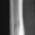
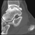
The types of imaging studies performed for patients with suspected stress fractures run the entire gamut including radiography, computed tomography (CT), nuclear imaging, and MRI. Ultrasound does not have a place in the diagnostic armamentarium for these conditions. Radiography is most often the initial imaging study performed. Unfortunately, it is not the most sensitive tool that we have for establishing a diagnosis in the early stages. Figure 11-4 shows the gamut of imaging and the points in time at which they are likely to yield positive results. The radiographic appearance depends on the time sequence between the onset of injury and the radiographic examination, as well as whether or not the patient continued participation in the activity. Mulligan described the earliest radiographic change as a “graying” or indistinctness of the cortex that he believed most likely represented localized bone or periosteal edema. Occasionally, in the early phase, the fracture in the shaft of a long bone may appear as a lucency through the cortex without any evidence of periosteal reaction or callus ( Figure 11-5 ). In cancellous bone, such as the calcaneus, the first manifestation may be a linear area of sclerosis that is frequently oriented perpendicular to the trabeculae ( Figure 11-6 ). Once healing begins, solid or thick lamellar periosteal reaction occurs on both the endosteal as well as the periosteal surface, confined to a small area of the cortex ( Figure 11-7 ). This fusiform area of reactive bone is typically smaller than that encountered in an osteoid osteoma. In addition, stress fractures typically involve only one of the cortical surfaces as opposed to the entire circumference of the bone, as in the case of sclerosing osteomyelitis. As periosteal reaction thickens, any fracture line that may have been present disappears.


