Tip of feeding tube should be located beyond stomach (distal duodenum or jejunum)
• Nasogastric tubes
Large-bore, moderately stiff
Used for temporary bowel decompression
Tip placed in pylorus can cause outlet obstruction
• Gastrostomy and jejunostomy tubes
Balloon-tipped catheters should not be placed into small bowel (may obstruct lumen)
Small amount of free air after placement is common and usually does not require intervention
IMAGING
• Malposition is most frequent complication of feeding tubes
Can be visualized on chest or abdominal radiograph
Auscultation over abdomen is not reliable method for confirming proper tube placement
CLINICAL ISSUES
• 1-3% of feeding tubes enter tracheobronchial tree
Anywhere from trachea to pleural space
Can perforate lung with significant morbidity and mortality
• Tube may penetrate esophagus or duodenum with fatal results
Often through diverticula (e.g., Zenker), due to thin wall
• High-risk patients
Altered mental status
Absent gag reflex
Multiple or repetitive insertion attempts
• Treatment
Reposition feeding tube if in incorrect location
Perforation of lung or bowel may require surgery
(Left) Esophagram shows a retroesophageal collection of gas and contrast medium resulting from perforation of a Zenker diverticulum by attempted placement of a feeding tube whose track runs parallel to the proximal esophagus.
(Right) Chest radiograph shows a feeding tube that has entered the right bronchus and perforated the lung though a lower lobe bronchus. The tip lies in the pleural space, a procedural complication that may be fatal, especially if food is given through the tube.
(Left) Frontal radiograph shows the peculiar course of the feeding tube with abrupt upper deviation of its distal portion. CT showed that the tube had perforated the duodenum and had been advanced with its wire in place.
(Right) Axial CECT shows a feeding gastrostomy tube entering the stomach. The balloon tip of the tube has migrated into the jejunum where it is partially occluding its lumen.
TERMINOLOGY
Definitions
• Patient injury caused by improper feeding tube placement
• Feeding tubes
Small, soft enteric tubes
Some with flexible metallic tips
Used for feeding chronically ill patients
Can be used for long periods of time
• Nasogastric tubes
Large-bore, moderately stiff
Used for temporary bowel decompression or fluid sampling
Only gold members can continue reading. Log In or Register to continue


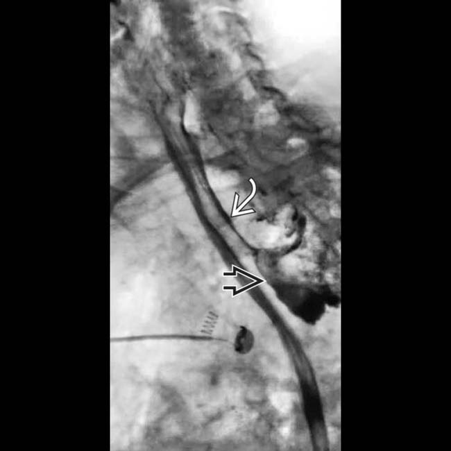
 resulting from perforation of a Zenker diverticulum by attempted placement of a feeding tube whose track
  runs parallel to the proximal esophagus.
 that has entered the right bronchus and perforated the lung though a lower lobe bronchus. The tip
  lies in the pleural space, a procedural complication that may be fatal, especially if food is given through the tube.
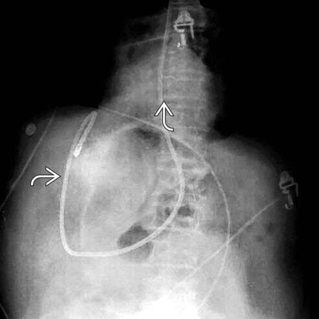
 with abrupt upper deviation of its distal portion. CT showed that the tube had perforated the duodenum and had been advanced with its wire in place.
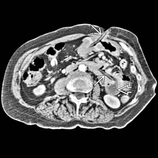
 entering the stomach. The balloon tip of the tube
  has migrated into the jejunum where it is partially occluding its lumen.


