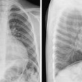
6 Identification, Clinical History, and Radiographic Techniques Before starting analysis of the radiographic findings, one should ascertain (1) the identification of the patient, including the name, medical record number, age, and gender; (2) the clinical information; (3) the right and left sides of the patient on the radiograph; and (4) the radiographic projection and exposure. The patient’s identification should always be checked every time the radiographs are reviewed. Putting an incorrect identification on a radiograph or taking radiographs from the wrong patient is a serious mistake that should be avoided, although it does happen. Recognition of incorrect labeling is easy when previous studies are available for comparison. The patient’s age, gender, and clinical information may provide clues in discovering incorrect identifications. The age of the patient should also be taken into consideration in radiographic interpretation, as normal radiographic findings vary with age. The clinical information given in the request for radiographs may include the clinical diagnosis, the patient’s major symptoms and signs, and the medical or surgical interventions. The radiographic findings at first should be reviewed blindly to avoid a potential bias, and then with the clinical information taken into consideration. The radiographic findings should be assessed as to whether or not they are in accordance with the given clinical information, the severity of the radiographic manifestations, and the patient’s diagnosis or condition. The findings should also be reviewed for any additional unrecognized or unsuspected features. Incorrect labeling of the right/left side of the chest is rare. But there are a few pitfall conditions that may lead the radiographic technologist to make this mistake. For example, at our hospital we were unable to place in the supine position a newborn with a meningomyelocele, so all radiographs were taken in the prone position. But the radiographs were incorrectly labeled with a side marker in the same way as for patients whose radiographs are routinely obtained in a supine position. We also had a patient with situs inversus and dextrocardia on whose radiograph the side of the chest was initially labeled with a proper side marker but later it was replaced with an incorrect marker when the technologist thought that there had been a mistake. Chest radiographs are usually obtained with the patient upright and in deep inspiration, using a posteroanterior projection for a frontal view and a right-to-left projection for a lateral view. These projections allow full expansions of the lungs for optimal assessment and minimize magnification of the cardiac silhouette. There are occasions when these standard projections are not possible. In particular, newborns and critically ill patients in the intensive care unit require imaging in a supine position, which is routinely performed using an anteroposterior projection. A large cardiac silhouette and indistinct cardiac borders result from both supine positioning of the patient and the anteroposterior projection (Fig. 6.1). An oblique radiographic projection to the right or left is not uncommon in pediatric patients. The configuration and size of the cardiovascular silhouette may appear significantly different in oblique views (Fig. 6.2). An oblique projection can be easily identified by comparing the positions of the anterior tips of the bony ribs. When a frontal chest radiograph is obtained in a left anterior or right posterior oblique view, the tips of the right ribs are aligned far outward along the lateral boundary of the right thorax, whereas the tips of the left ribs are projected inward (Fig. 6.2, left panel). When a frontal chest radiograph is obtained in the right anterior or left posterior oblique view, the tips of the left ribs are aligned far outward, whereas the tips of the right ribs are projected inward (Fig. 6.2, right panel). When there is levocardia, the cardiac silhouette is smaller in the left anterior or right posterior oblique view than in the straight frontal view because the short axis of the heart is projected toward the radiographic screen in the former. In contrast, the cardiac silhouette is larger in the right anterior or left posterior oblique view because the long axis of the heart is projected toward the screen. When there is dextrocardia, this relationship between size and obliquity is reversed (Fig. 6.3).
 Identification
 Clinical Information
 Right/Left Sides of the Patient on Radiographs
 Radiographic Projection and Exposure
Stay updated, free articles. Join our Telegram channel

Full access? Get Clinical Tree


