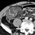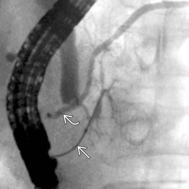
 and accessory
  pancreatic ducts do not communicate. This results embryologically from failure of fusion of the ducts between the dorsal and ventral pancreatic anlagen.
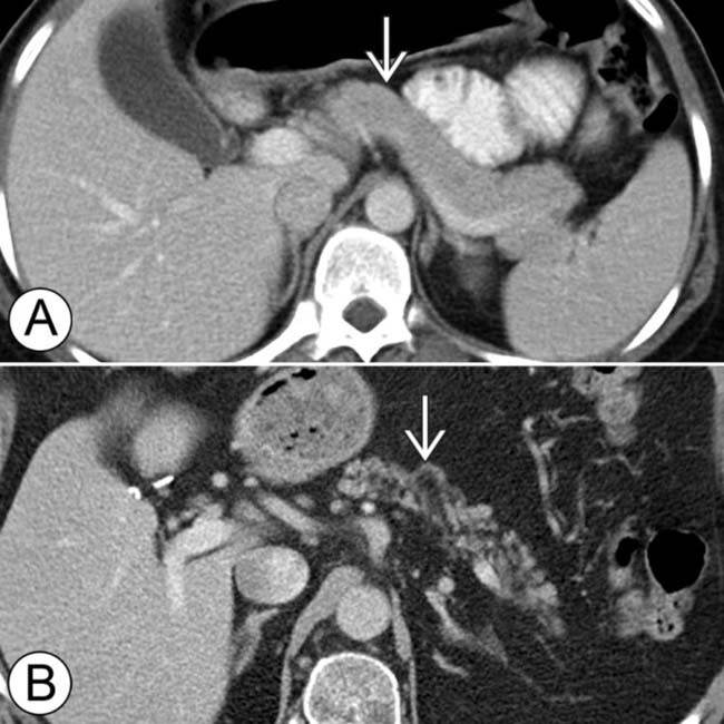
 . The top image (A) is from a 30-year-old woman and the bottom image (B) is from a 78-year-old man. With aging, the pancreas decreases in size with increased fatty lobulation. Small calcifications and mild ductal dilatation may also be seen.
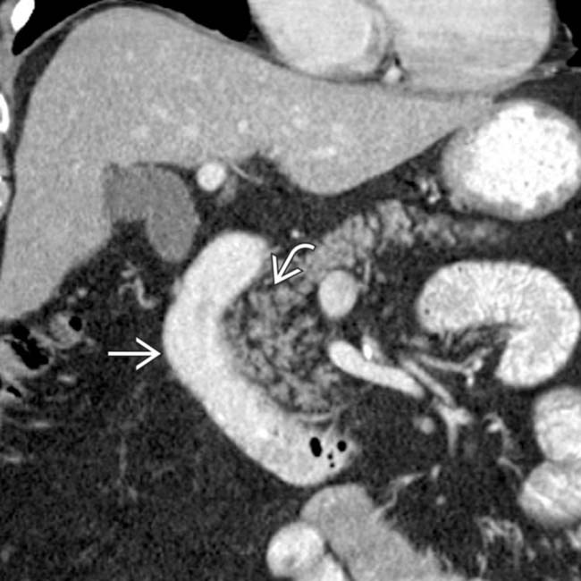
 to the 2nd portion of the duodenum
  .
.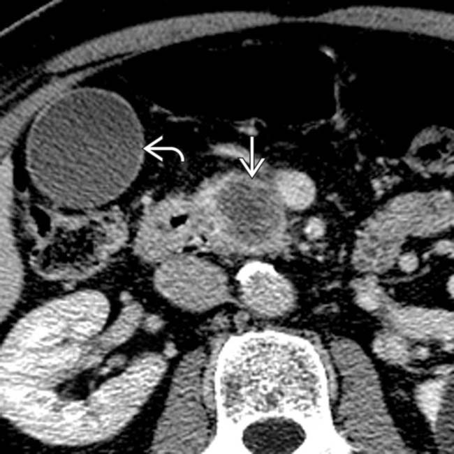
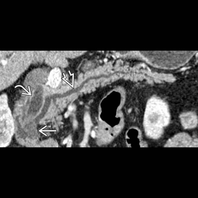
 in the head of the pancreas and a distended gallbladder
  .
.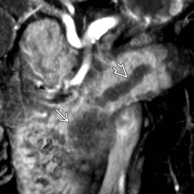
 interrupted as it enters the hypodense mass
  , a typical presentation of pancreatic ductal carcinoma.
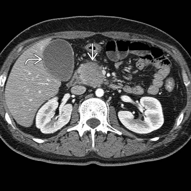
 in the head of the pancreas causing biliary obstruction and dilation of the gallbladder
  .
.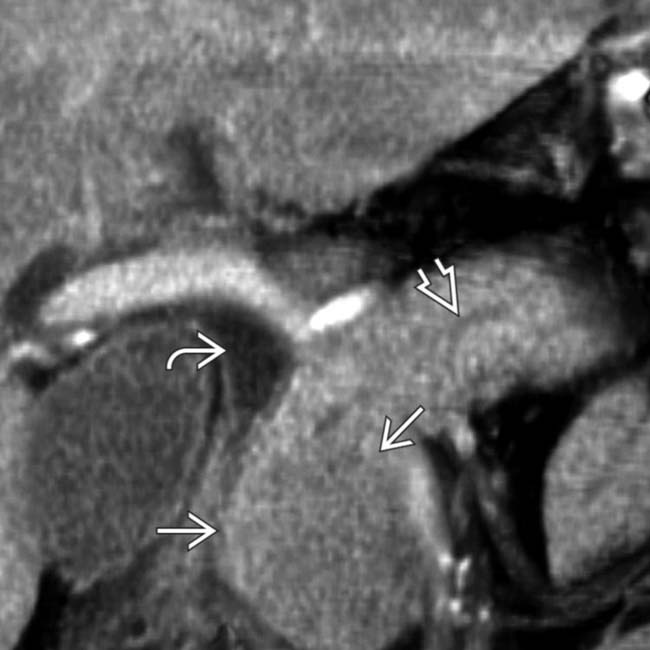
 causing partial obstruction of the bile duct
  , while the pancreatic duct
  is only mildly dilated. Further evaluation, including biopsy, confirmed a diagnosis of autoimmune (IgG4-related) pancreatitis.
 and pancreatic duct
  due to a small hypodense ampullary carcinoma
  .
.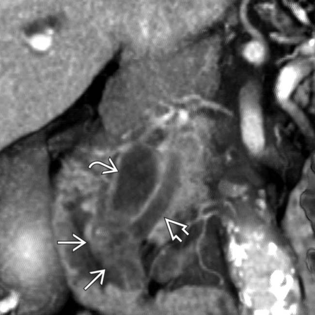
 , pancreatic duct
  , and ampullary tumor
  .
.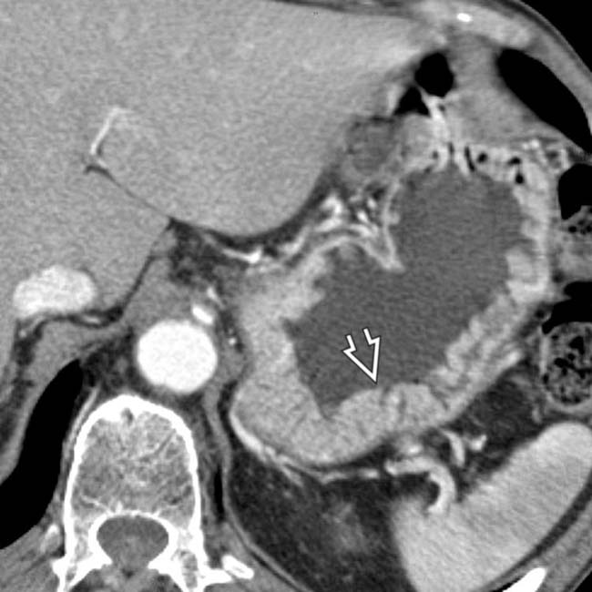
 .
.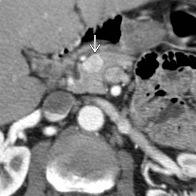
 in the pancreatic head that proved to be a gastrinoma (1 type of pancreatic endocrine tumor) that was responsible for this patient’s Zollinger-Ellison syndrome.
Stay updated, free articles. Join our Telegram channel

Full access? Get Clinical Tree


