The integration of imaging characteristics and genomic data has started a new trend in approach toward management of glioblastoma (GBM). Many ongoing studies are investigating imaging phenotypical signatures that could explain more about the behavior of GBM and its outcome. The discovery of biomarkers has played an adjuvant role in treating and predicting the outcome of patients with GBM. Discovering these imaging phenotypical signatures and dysregulated pathways/genes is needed and required to engineer treatment based on specific GBM manifestations. Characterizing these parameters will establish well-defined criteria so researchers can build on the treatment of GBM through personal medicine.
Key points
- •
The integration of imaging characteristics and genomic data has started a new trend in the approach toward management of glioblastoma multiforme (GBM).
- •
Recently many ongoing studies are investigating imaging phenotypical signatures that could explain more about the behavior of the GBM and its outcome decisively.
- •
The discovery of biomarkers has played an adjuvant role in treating and predicting the outcome of patients with GBM.
- •
Discovering these imaging phenotypical signatures and dysregulated pathways/genes is the need of the hour and is a prerequisite to engineer treatment based on specific GBM manifestations.
- •
Characterizing these parameters will lay terra firma and establish well-defined criteria so researchers can build on and revolutionize the treatment of GBM through personal medicine.
Introduction
Among several forms of brain malignancies, glioblastoma (GBM) is the most common malignant brain neoplasm, accounting for 29.5% of all primary brain tumors. In the United States alone more than 10,000 patients are newly diagnosed with GBM every year. Current treatment is aggressive multimodal therapy with targeted surgical resection followed by radiation therapy and temozolomide. Despite the prevailing standards of treatment, GBM still has an appalling prognosis with an overall median duration of survival ranging from 12.2 to 15.9 months.
The largest cause of failure in GBM therapy is attributed to the inherent heterogeneity existing in intratumoral tissues. This heterogeneity, in turn, leads to a varying response to treatment and poor patient prognosis. Using molecular techniques to understand the underlying cause for GBM would aid greatly in tackling this multifaceted tumor. Relying merely on histopathologic evidence would be detrimental to successful treatment of GBM, and a dependable genetic characteristic needs to be identified to improve accuracy and ensure effective therapy. The genetic heterogeneity of the tumor translates to different expression patterns within the neoplasm, which influences the magnetic resonance (MR) image. To develop personalized targeted therapy it is critical to identify these patterns of genetic alterations in GBM.
Given the nascent nature of imaging genomics, it still does not have a standard definition. It can be thought of as an integration of medical imaging data and -omics data, which represents chiefly MR imaging on one hand and gene, protein, or metabolite expressions on the other. Thus, imaging genomics has a lot of potential in identifying morphologic and expressional profiles of the tumor with a holistic view while drawing parallels with the visible phenotypical results obtained from MR images.
Microarray techniques
With the advent of breakthrough molecular biology techniques, the microarray has gained importance in its ability to detect mRNA expression levels and analyze whole genomes and individual genes. Using microarrays can help discover new targets of therapy for treatment of GBM, and there is currently an ongoing effort to find such targets. Micro-RNAs (mRNAs) are small-chain RNAs that do not code for any protein. Their mode of operation is different from regular RNAs in a way that they can regulate the expression of several target genes simultaneously. They carry the code to match the upstream 3′ UTR sequences of genes and can bind there to knockdown their expression. mRNAs have been used as a powerful gene-silencing tool to discover new pathways and determine protein interactions in a cell. This technique could be the answer to the problem of lack of targets for treating GBM. Using mRNAs to alter the expression of oncogenes and tumor suppressors seems a promising strategy to find novel therapy by regulating proliferation, apoptosis, neovascularization, invasion, and migration.
All said and done, microarray does have its shortcomings. The source of tissues for running such microarray assays is the resected tumor (or biopsy) from patients. These tissues are not only composed of tumor tissue but also the adjoining healthy cells, stroma, and so forth. So the microarray is not a perfect representation of the gene profile of the tumor. Secondly, the accuracy of the microarray depends largely on the availability of a specific probe, varying sensitivity of multiple probes, among other parameters. The lack of specific probes can result in skewing of the outcome of the assay and paint a different picture of mRNA expression. Thirdly, mRNA expression measurements from different parts of the tumor only reflect the level of transcription, whereas measurement of protein levels would serve as more reliable data to assess the tumor’s subtype. Also, microarrays performed of different systems are coherent with different standards and cannot be compared directly, hence, mandating the need for normalization.
Molecular classification
Traditionally, GBM has been classified as primary and secondary: primary is composed of advance cancer with no evidence of low-grade lesions before diagnosis, and those tumors that have been detected at an earlier stage and subsequent evidence of progression (radiological or histopathologic) into a high-grade tumor was classified as a secondary tumor.
In recent years, based on genetic aberrations, Phillips and colleagues and Verhaak and colleagues independently classified GBM into differing subtypes. Phillips and colleagues emphasized the variations in expression levels of epidermal growth factor receptor ( EGFR ) and phosphatase and tensin homolog ( PTEN ). Accordingly, GBM was subdivided as proliferative ( PTEN loss and increased/normal EGFR ), proneural (normal PTEN and EGFR levels, notch pathway activation), and mesenchymal (increased neural stem cell markers, Akt activation, PTEN loss, and occasional EGFR activation). The proneural type of GBM had shown better survival rates in comparison with the mesenchymal and proliferative subgroups.
In a similar fashion, Verhaak and colleagues also classified GBM into 4 subtypes based on genetic variations in genes, such as tumor suppressor p53 ( TP53 ), EGFR , PTEN , isocitrate dehydrogenase-1 ( IDH1 ), neurofibromatosis type-1 ( NF1 ), and platelet-derived growth factor receptor A ( PDGFRA ). Classic GBM was predicted to have the longest survival when subject to vigorous treatment among all classes. Besides having common mutations in the TP53 gene, the proneural and mesenchymal subgroups differed in their genetic background indicated chiefly by variations in the PDGFRA , PTEN , IDH1 , and NF1 genes. Lastly, there also existed a neural subtype of GBM that commonly affected elderly patient groups; it shared common mutations with other subgroups with no specific gene dysregulation.
Biomarkers
The genetic aberrations or their varying phenotypic expression levels in tumor cells, blood, or urine can be used as a potential prognostic and diagnostic parameter, also called a biomarker. These biomarkers may reliably help in determining the prognosis, response to treatment, and survival and also allow identification of a resistance mechanism in patients with GBM undergoing treatment.
O-6-methylguanine-DNA Methyltransferase
The O-6-methylguanine-DNA methyltransferase (MGMT) gene is located at 10q26 and is responsible for DNA repair mechanisms in cells. This enzyme removes alkylating agents that are attached to the guanosine bases in DNA. At times, the expression of this enzyme is silenced because of the methylation of its promoter region. In such patients, not only is the DNA repair mechanism impaired but also treatment with alkylating agents is far more effective as the MGMT enzyme is absent, thus improving response to therapy. It has been shown clinically in a randomized phase III trial involving 206 subjects (hazard ratio = 0.45 and 95% confidence interval [CI] [0.32–0.61] P <.001) that patients who have an epigenetic silencing of this MGMT gene (by methylation) have responded better with temozolomide, radiotherapy, and showed a good prognosis. The median survival in the patients having the methylated MGMT gene with concomitant therapy with temozolomide and radiotherapy was 23.4 months as compared with normal individuals with active MGMT genes whose survival was only 15.3 months. MGMT methylation status was the first biomarker that indicated GBM prognosis and since then has been predominantly important in predicting the response to alkylating agents. However, it was not useful in aiding the diagnosis and designing treatment strategies for affected individuals.
Isocitrate Dehydrogenase–1
IDH is the rate-limiting enzyme that acts on the substrate isocitrate and converts it into α-ketoglutarate. This in turn protects the cell from oxidative damage as this reaction also reduces NADP to NADPH as a byproduct. Among the isoforms of IDH, the cytosolic IDH1 is of pertinence to GBM. IDH1 has been known to have mutations at the 132nd amino acid (arginine to histidine). This mutation increases its affinity to 2-oxaloglutarate and produces an oncometabolite: 2-hydroxyglutarate (2-HG). 2-HG in turn acts on hypoxia inducible factor 1α (HIF-1α) and hydroxylates it. An increase in HIF-1α activation is believed to be instrumental in progression of tumor in the case of GBM.
Janson and colleagues made a noteworthy clinical observation in 149 patients with GBM, of which 12% (n = 18) were found to have IDH1 mutations and showed increased overall survival (OS) of 31 months in patients who were IDH1 mutant versus 15 months in patients with normal IDH1. It is recently reported that the 5-year survival rate in patients with IDH1 mutations was 93%, which is significantly higher than 51% as seen in their IDH1-negative counterparts. Thus, these findings strongly imply that improved outcome, reduced aggressiveness, and better prognosis in high-grade glioma cases have been associated with mutations in the IDH1 gene. In recent years, studies highlight the potential application of IDH1 in determining therapy outcome. Song Tao and colleagues reported that the median progression-free survival rate for patients with both IDH1 and MGMT mutations in response to temozolomide treatment was 13.4 months, whereas those who had only IDH1 mutation showed a median survival rate of 10.2 months. Patients who lack any mutations in the aforementioned biomarkers had the least median survival rate of 6.1 months.
Deletion of 1p/19q (Loss of Heterozygosity)
Deletion of 1p/19q (loss of heterozygosity [LOH]) has been found to be a significant biomarker in cases of oligodendromas. The 1p/19q deletions in GBM are found to be uncommon (<10%) in diffuse astrocytic gliomas. Contrastingly, the most common genetic mutations (40%–90%) in the oligodendroglioma component of GBM is 1p and 19q codeletion, which is associated with a favorable response to chemotherapy, radiation, and survival. These tumors with 1p/19q codeletion have a greater propensity to occur in the frontal lobe as compared with insular tumors that rarely show deletion or alteration in neither 1p nor 19q. In a trial study by the European Organization for Research and Treatment of Cancer/National Cancer Information Center, 360 subjects with GBM were studied for their differential response to chemo-radiotherapy with temozolomide, whereby the particular focus was on the presence of an oligodendromalike component (GBM-O). It was reported that 15% of the total population were positive for the presence of GBM-O. Another key finding of this study was that this 15% of patients with GBM-O had an ominous outcome. But codeletion of 1p/19q was found in only 1 case. There is some ambiguity associated with the correlation of 1p/19q codeletions and prognosis of GBM. In one study performed by Hill and colleagues it was reported that there was an improvement in prognosis of GBM cases with 1p/19q deletion. Conversely, in another study, despite the presence of an oligodendroma component there was no apparent improvement in prognosis. Jesionek-Kupnicka and colleagues, however, reported that there was no reciprocity between LOH of 1p/19q codeletion and patient survival.
A study conducted by Homma and colleagues of 209 patients with GBM sought to find parallels between 1p deletions and prognosis. Interestingly, the results showed that patients (21% of population) with 1p LOH had significantly longer duration of survival (13.1 ± 10.8 months) than the patients without the LOH (9.6 ± 7.4 months; P <.05). These results were conclusive even after further adjustments were made for age and sex in a multivariate analysis; survival in patients with LOH of 1p had a hazard ratio of 0.7 (with 95% CI; 0.5–1.0).
Epidermal Growth Factor Receptor
EGFR is a tyrosine kinase–linked receptor that is present on epidermal cells. It dimerizes on activation to switch on signaling and increase phosphoinositide 3 kinase (PI3K) and subsequently PTEN . This aids in increasing neovascularization, cell proliferation, and survival in tumors. It is observed that EGFR is overexpressed in about 50% to 60% of patients with GBM. The most common variant of EGFR seen in these cases is the EGFRvIII (24%–67%) that activates the EGFR-PI3K pathway constitutively. A genetic sense-mutation leading to the deletion of exons 2 to 7 results in premature truncation of the extracellular ligand binding domain of EGFR, hence, resulting in expression of EGFRvIII.
Several studies have been conducted to screen for correlations of wild-type-EGFR (wtEGFR) as a prognostic factor in patients with GBM; and results showed wtEGFR as not an independent favorable prognostic factor. However, these results depict only a skewed image of reality as one study had a limited sample size; another study used ill-defined resection of surgical cavity and variations in postoperative treatment. Although tumor invasion seems to be more profound in the EGFRvIII as compared with wtEGFR, EGFRvIII expression levels have not been used as an independent factor for determining prognosis in patients with GBM. A heterogeneous study by Shinojima and colleagues conveyed EGFRvIII overexpression in the presence of EGFR amplification as a potent indicator of negative prognosis. However, a homogenous study by Heimberger and colleagues concluded that EGFRvIII is as an independent negative prognostic predictor in patients with GBM who survived greater than 1 year.
Besides being a potential negative prognostic factor of GBM, EGFR can also be a potential target for therapy, as a reported 97% of patients with primary classic GBM show EGFR amplification. So, use of an EGFR inhibitor could be useful in treating this subtype of GBM. Hence, therapy can be tailored better by determining the EGFR amplification/overexpression in patients.
Vascular Endothelial Growth Factor
VEGF is an important proangiogenic factor that is highly regulated by HIF-1α. Hypoxia is commonly seen in tumors whereby the core is starved of oxygen. VEGF mediates this response to hypoxia by inducing growth of new blood vessels, which have a higher permeability, thus, causing local edema. Nearly 64.1% of GBM cases have been found to have increased VEGF. Further, a study that was conducted by Carlson and colleagues in 2007 whereby they studied the relationship between VEGF expression and edema revealed that VEGF expression levels correlated well with tumor grade. GBM showed a 4-fold increase in VEGF expression when compared with grade 3 tumors. They outlined 2 groups of patients: no edema and little or abundant edema based on imaging characteristics. Interestingly, the group that had no edema showed no VEGF overexpression; on the other hand, the group with little or abundant edema showed higher VEGF levels, which correlated with survival.
Subsequently, Pope and colleagues reported another study in 2008 that considered completely enhancing (CE) and incompletely enhancing (IE) tumors of GBM based on MR images. Gene analyses were then conducted on these two groups, and the disparity in gene expression was studied. CE tumors were characterized by an increase in VEGF among other genes when compared with IE tumors, and a conclusion was drawn that targeting VEGF in these cases could be very important to improve response to therapy. VEGF is not a solitary factor that orchestrates the neovascularization and progression of tumor in GBM. It works in conjunction with other proteins, such as interleukin-8 and matrix metalloproteinase 7, to further tumor progression and cell survival. This finding is shown by the clinical study whereby monotherapy using bevacizumab (a VEGF-targeting monoclonal antibody) has not been very successful in prolonging tumor-free survival and patient survival. A global view of GBM can be obtained using MR imaging to track edema in tumors, and correlations can be made to monitor effectiveness of therapy and predict prognosis.
Quantitative analysis of imaging genomics
Quantitative measurement parameters have been recently studied for possible clues of expected survivability in patients with GBM, which could not have been accomplished without the aid of the qualitative imaging analyses performed over the last decade. A pioneering study using volumetric MR imaging by Zinn and colleagues in conjunction with molecular biology techniques show some very interesting results. The study focused on analysis of gene expression in high versus low FLAIR patients. In the ingenuity pathway analysis of the 53 genes that were screened, 29 genes were shown to have a significant part in cancer, whereas another 20 genes were shown to play a role in cell migration and invasion and 16 genes played a common role in cancer and invasion. However, the gene periostin (POSTN) seemed to have the largest increase in expression in high versus low fluid-attenuated inversion recovery (FLAIR) patients (about a 7.5–mean fold increase). Furthermore, reports stated that POSTN was upregulated in the mesenchymal versus proneural subtype ( P <.0001). POSTN expression has been shown to increase under TWIST-1 induction and modulate the migratory response in vitro. The switch from the epithelial type to the mesenchymal type (called EMT) is one of the critical ways of cell invasion and treatment failure, which implies POSTN in some way responsible for influencing the migratory character of the tumor in vivo, which could explain the outcome of Zinn and colleagues’ study. The classic method of distinguishing patients with GBM into proneural and mesenchymal is a relatively poor indicator of patient survival with a P value of .202. However, when POSTN expression levels were considered as an independent determining factor for survival, its correlation with survival was found to be statistically significant ( P = .03) in a Cox proportional hazards model. There have been parallels showing the existence of overlaps between tumor invasiveness and tumor resistance; because POSTN is a viable marker for tumor invasiveness, it could also serve as a marker for predicting response to therapy in patients with GBM.
In 2012 Zinn and colleagues published another study using quantitative analysis and built a preoperative prognostic predictive model in patients with GBM. The variables of their predictive models were (1) volume of the tumor, (2) patient age, and (3) Karnofsky performance status (KPS); hence, they presented a new clinically applicable VAK classification. The VAK classification used a simple 3-point scoring system that would allow bisection of patients with GBM into VAK-A (score of 0 or 1) and VAK-B (score of 2 or 3). It was reported that patients with larger tumor volumes showed decreased survival compared with patients with smaller lesions ( P = .14, survival 12.0 vs 18.5 months) when the median cutoff (30,000 mm 3 ) was considered as previously described in the literature. Although KPS and age did not hold much significance independently, when combined with the tumor volume parameter, the prognostic value of the aforementioned variables was enhanced (age, KPS, and tumor volume) and dividing the patient population was aided, with P = .007, survival 12 versus 20 months (VAK-B vs VAK-A). The patient data from The Cancer Genome Atlas (TCGA) and Rembrandt’s database were used in validating the VAK model; the resulting VAK classification was more significant in predicting patient survival when corrected for age and KPS. Such innovative methods for predicting patient prognosis preoperatively is made possible because of the ability to accurately quantify tumor volume in lesions using MR imaging. The investigators also furthered their study by examining variations in patterns of genetic expression (TP53 and MGMT methylation) and incorporated it into the VAK classification with better correlation to patient survival.
An interesting recent study was conducted by Colen and colleagues involving a sex-specific prediction of cell death by alternative mechanisms of apoptotic pathways using imaging genomics. Female patients were found to have smaller volumes of necrosis when compared with their male counterparts (6821 vs 11,050 mm 3 , P = .03). Female patients (n = 30) who showed a lower volume of necrosis on imaging had significantly higher benefit of survival of 11.5 months when compared with the female patients with higher necrosis volumes ( P = .01). Male patients (n = 69) of high and low necrosis groups did not show any change in survival. Although male patients with low necrosis volumes did not show any significant benefit in survival when compared with female patients with low necrosis volumes (n = 50, P = .9), the male patients with high necrosis volumes showed significantly higher survival benefit (8 months) when compared with female patients with high necrosis volumes (14.5 vs 6.5 months; P = .01, n = 49).
In an effort to understand this phenomenon, the investigators examined the mechanisms of apoptosis in both groups and arrived at the conclusion that the TP53-mediated apoptosis in male patients was the reason for increased necrosis, whereas the female patients had a differing mechanism of MYC oncogene dysregulation mediating necrosis. This finding opens a new avenue of potential therapeutic targets to treat sex-specific and sex-nonspecific mediation of necrosis in patients with GBM.
Gevaert and colleagues conducted a study to extract quantitative features from MR images of the regions of interest (ROI) (enhancing necrotic zones and the surrounding peritumoral edematous areas) that characterize the radiographic phenotype of GBM lesions to create a radiogenomic map that correlates quantitative imaging features with molecular data.
A part of their study concentrated on correlating robust quantitative image features with survivability (OS and progression-free survival). Interestingly, 3 noteworthy correlations for enhancement image features were reported ( P <.05): (1) OS was shown to highly correlate with variance of the radial distance signal of the enhancement of the ROI feature ( P = .017; hazard ratio = 0.67). This feature defines the irregularity of the border of the ROI, and a smaller value would imply good prognosis. (2) The sharpness of the edge was also shown to correlate with OS, the smoother indicating better prognosis ( P = .023; hazard ratio = 1.43). (3) In a similar fashion, a lesser blurriness of the edge was reported to have correlated with progression-free survival ( P = .028; hazard ratio = 0.48). By this study the investigators highlighted the emphasis of quantitative image analysis in GBM and reported significant correlations with survival.
A study by Naeini and colleagues demonstrates a simple method to potentially predict the mesenchymal (MES) subtype of GBM in patients and their survival using various ratios of quantified zones in MR images. The quantifiable parameters are (1) volume of contrast enhancement, (2) volume of central necrosis, (3) hyperintensity of T2/FLAIR, and (4) ratio of T2/FLAIR hyperintense volume to the volume of contrast enhancement and necrosis.
Among the different parameters that were calculated, the one the authors are interested in is the ratio of T2/FLAIR hyperintense volume to contrast enhancing and necrosis volume. A ratio of more than 3 had a sensitivity of 71% and a specificity of 100% for predicting the MES subtype. The investigators then classified patients based on the T2/FLAIR hyperintense volume to contrast enhancing and necrosis volume ratio into long (ratio >3.0) and short (ratio <3.0) OS groups ( P = .0064). Then a multivariate Cox proportional hazards ratio analysis was performed to confirm that the ratio is able to independently classify the patients into short- and long-term survival groups when age and KPS are taken into consideration (Cox regression; ratio >3.0 vs ratio <3.0 covariate, P = .05; age covariate, P = .9617; KPS covariate = 0.1410). Thus, this study shows that the ratio of T2/FLAIR hyperintense volume to contrast enhancing and necrosis volume could be potentially a straightforward and influential marker for predicting the MES subtype of GBM from other phenotypes and their survival.
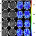
Hirai and colleagues in 2008 came up with an unconventional yet radical study (n = 49) that correlated high-grade gliomas (GBM and astrocytoma) and prognosis to MR imaging features using relative cerebral blood volume (rCBV). The investigators used receiver operating characteristic analysis to establish an rCBV cutoff of 2.3, which showed a sensitivity of 98% and specificity of 98% (positive predictive value = 66%; negative predictive value = 90%). When the investigators correlated the maximum rCBV and survival time, they found the 2-year survival to be significantly lower in patients with GBM (4 of 31 individuals) with high rCBV (≥2.3) when compared with patients with astrocytoma (16 of 18 individuals; P <.001). In patients with high-grade gliomas exhibiting low rCBV (18 of 27), the 2-year survival was shown to be significantly higher when compared with patients with a high rCBV (2 of 22; P <.001).
In 2013 Jain and colleagues performed a study along similar lines as Hirai and colleagues whereby they used perfusion CT to measure rCBV and permeability surface area-product (PS) in patients with high-grade glioma retrospectively and correlated it with patient OS. When they concentrated on grade 4 gliomas, the rCBV was statistically significant ( P = .019) between high and low rCBV with a 2-year survival rate being 24% (median OS = 14.2 months) for high rCBV when compared with low being 86% (median OS not reached). The investigators reported that contrastingly when only PS was considered for the correlation, there was no statistical relationship between PS (high vs low) and OS. However, the median OS was significantly higher in the high rCBV + PS group when compared with the low rCBV + PS group (39.5 months vs 13.6 months; P = .004). Thus, rCBV and PS together served as a much better indicator of patient survival than the two parameters considered independently. This finding promises the capacity of the use of vascular imaging parameters as prognostic biomarkers in high-grade gliomas.
Related posts:
 New State-of-the-Art and Cutting-Edge Advances in Brain Tumor Imaging
 Dynamic Susceptibility Contrast MR Imaging in Glioma
 Response Assessment in Neuro-Oncology Criteria and Clinical Endpoints
Perfusion Imaging in Neuro-Oncology
Shedding Light on the 2016 World Health Organization Classification of Tumors of the Central Nervous System in the Era of Radiomics and Radiogenomics
Adult Brain Tumors
Response Assessment in Neuro-Oncology Criteria and Clinical Endpoints
Perfusion Imaging in Neuro-Oncology
Shedding Light on the 2016 World Health Organization Classification of Tumors of the Central Nervous System in the Era of Radiomics and Radiogenomics
Adult Brain Tumors
Stay updated, free articles. Join our Telegram channel

Full access? Get Clinical Tree


