Imaging Techniques

Chapter 130


Imaging Techniques


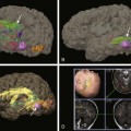
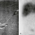
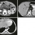
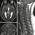
Pediatric musculoskeletal radiology is a broad field that requires an understanding of normal growth and developmental variations, fracture patterns unique to the immature skeleton, skeletal dysplasias, and knowledge of unique tumor and tumor-like conditions. Advanced imaging has improved our ability to arrive at a precise diagnosis. However, it also has created the need for additional expertise within the field of pediatric imaging to learn how to properly use these tools to arrive at a diagnosis and provide information beyond that of the humble radiograph. This section will cover the spectrum of pediatric musculoskeletal disorders from a multimodality imaging approach. Key images are provided in the printed version and an expanded, comprehensive image library is provided in the electronic version.


Imaging Technique Overview


Radiography


Radiography remains the initial tool for the evaluation of acute trauma, inflammatory arthritis, infection, suspected primary bone neoplasms, and skeletal dysplasias.


In the setting of acute trauma, nonarticular long bones should be imaged with at least two views (frontal and lateral). Osteoarticular regions should be imaged with three views (frontal, lateral, and oblique). Dedicated imaging of the digits is preferred rather than general imaging of an entire hand or foot when a patient has a single symptomatic digit. The radiograph should be the initial screening tool before computed tomography (CT) or magnetic resonance imaging (MRI) is performed in the setting of acute injuries. For alignment disorders, including scoliosis and foot deformities, weight-bearing views should be obtained routinely. In cases of suspected child abuse, a dedicated skeletal survey should be performed. Bone scintigraphy and MRI are complementary tools in the evaluation of child abuse, but they may miss the classic metaphyseal corner fracture.1


In the setting of infection, radiography should be performed before advanced imaging, although a normal radiograph should not preclude referral for MRI for suspected acute osteomyelitis. Radiographs are helpful for initial screening before the use of MRI to ensure that symptoms of suspected infection are not a result of an underlying fracture or primary bone neoplasm.


For primary bone neoplasms, radiographs are key to determining coverage by MRI; they also complement the MRI diagnosis. Radiographs may help with the final diagnosis because the matrix and pattern of bone destruction are better delineated by radiography than by MRI.


Ultrasonography


Musculoskeletal ultrasound has three primary roles: evaluation of dysplasias, of soft tissue masses, and of pyogenic and nonpyogenic arthritis.


Sonographic evaluation of dysplasias includes developmental dysplasia of the hip, glenohumeral dysplasia related to brachial plexopathy, and selected congenital foot deformities. For developmental dysplasia of the hip, the optimal time of imaging is before the capital femoral epiphyseal ossification center appears, which usually is at 4 to 6 months of age and younger. Sonography can image through cartilage but not bone. Sonography can evaluate the relationship of the humeral head with respect to the glenoid in the setting of suspected glenohumeral dysplasia and subsequently can be used to calculate a glenoid version angle. Sonography also is useful for certain congenital foot disorders and can determine the relationship between the nonossified navicular and talus in the setting of congenital vertical talus.

Stay updated, free articles. Join our Telegram channel

Dec 20, 2015 | Posted by in PEDIATRIC IMAGING | Comments Off on Imaging Techniques

Full access? Get Clinical Tree

Get Clinical Tree app for offline access