Key Facts
- •
The terms osteonecrosis (ON) and avascular necrosis (AVN) are usually used interchangeably. The term infarct usually refers to identical changes occurring in the marrow cavity rather than in the subchondral bone.
- •
One theory indicates that bone marrow edema may lead to increased intramedullary pressure and osteonecrosis.
- •
The “crescent sign” on radiographs is a subchondral fracture resulting from bone resorption. It is characteristic of ON but is not an early finding.
- •
Magnetic resonance imaging (MRI) is the most sensitive and specific test for the diagnosis of ON (or infarct), but false-negative cases have been reported.
- •
The margin of an osteonecrotic segment is well defined. A low signal rim with an inner high signal band on T2-weighted sequences, “the double line sign,” is a characteristic MRI feature of ON.
- •
If there is a clinical question of ON, radiographs should be done including a “frog leg” lateral view. If these are negative on one or both sides and treatment is considered, MRI without contrast is indicated. If MRI and radiographs are normal and clinical suspicion of early ON remains, contrast-enhanced MRI examination or bone scan could be done.
- •
Bone marrow edema may be seen in patients with ON, transient osteoporosis, or insufficiency fracture. The differentiation between these conditions may be difficult on imaging studies early in their course. Clinical correlation and follow-up studies may be necessary.
- •
Spontaneous osteonecrosis of the knee (SONK) affects older women, is abrupt in onset, and usually affects the medial femoral condyle. It may be due to a subchondral insufficiency fracture.
OSTEONECROSIS
Avascular necrosis (AVN) is also known as ischemic necrosis, osteonecrosis , or aseptic necrosis . All these terms refer to bone death resulting from insufficient blood supply to the subchondral bone. The term infarction is usually applied to the identical changes occurring in the marrow cavity rather than in the subchondral bone.
Osteonecrosis may be posttraumatic occurring secondary to femoral neck fractures, hip dislocations, or the forcible reduction of the hip in patients with developmental dysplasia. In these cases, the trauma interrupts the medial and lateral femoral circumflex arteries, causing osteonecrosis. This chapter reviews nontraumatic osteonecrosis.
Osteonecrosis is the underlying reason for 5% to 12% of total hip replacements.
Causes
Osteonecrosis (AVN) has been described to result from a host of different abnormalities with the common end point being mechanical failure followed by collapse of the articular surface. In one large series, 90% of cases occurred in patients who were alcohol abusers or were on corticosteroids.
The minimum dose of corticosteroids necessary to result in osteonecrosis is thought to be equivalent to 4000 mg for a period of 3 months, although this complication has been reported with even low-dose, short courses of treatment.
Other etiologies of osteonecrosis include hemoglobinopathies, in particular sickle cell and Sickle-C disease, marrow packing disorders (Gaucher’s disease), and barotrauma ( Box 16-1 ). In approximately 5% of patients with osteonecrosis, the cause is not apparent (idiopathic), but in many of these patients the cause is unrecognized alcohol abuse. There is an increased risk of osteonecrosis with as little as 400 mL of alcohol per week, which is the equivalent of approximately 20 beers.
Trauma
Vasculitis
Corticosteroids
Sickle cell disease, Sickle C
Barotrauma (nitrogen emboli)
Gaucher’s disease
Alcoholism (multifactorial)
The pathophysiology of osteonecrosis is unknown and multiple theories have been proposed. Ficat and others believe that bone is a fixed compartment and increased intraosseous pressure resulting from ischemia and marrow edema resulting in osteonecrosis. The increased pressure may be secondary to venous abnormalities, which can be seen in sickle cell disease secondary to sickling in the intraosseous veins, and venous obstruction could be caused by nitrogen bubbles in dysbaric osteonecrosis. Extravascular obstruction can occur secondary to marrow packing in Gaucher’s disease. This compartment theory forms the basis for core decompression as treatment, in which a cylindric core is removed from the femoral head and neck to decrease intraosseous pressure. Jones believes that osteonecrosis is multifactorial and that abnormal fat metabolism with stasis, hypercoagulability, and endothelial damage by free fatty acids lead to fatty emboli and osteonecrosis in patients who abuse alcohol or are on corticosteroids.
Pathologic Findings
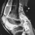
Sweet and Madewell divided the histologic appearance of osteonecrosis into four characteristic zones: a central area of cell death surrounded by an area of ischemia, an area of hyperemia, and an area of normal bone ( Figure 16-1 ). The “reactive zone” or interface is made up of the areas of hyperemia and ischemia. Histologically, the findings of ischemic necrosis are identical to those seen in bone infarction; the difference is based upon the location of the ischemia; when ischemia occurs in the subchondral bone it is referred to as osteonecrosis or AVN , and when it occurs in the shaft or metadiaphyseal region, the lesions are termed “bone infarcts” ( Figure 16-2 ).


Radiologic and histologic correlation reveals that initially normal findings are followed by bone resorption with a fibrous margin. Bone deposition at the margin or peripheral area of repair causes a sclerotic rim, giving the appearance of a “cyst like” lesion. Increased bone resorption results in macrofractures and eventually a visible crescent sign due to a subchondral fracture. This is followed by macrofracture with femoral head collapse and secondary osteoarthritis.
Standard radiography is insensitive for the early diagnosis of AVN and magnetic resonance imaging (MRI) is considered the most sensitive imaging study for diagnosis. MRI is occasionally negative, however. Koo and colleagues reported 28 hips (in 22 patients suspected clinically of having AVN) that had normal MRI examinations but had osteonecrosis diagnosed by angiography, scintigraphy, and intraosseous pressure measurements with biopsy. These patients were observed for 1 to 2 years. Eleven of the angiographically positive hips remained negative on MRI as did 14 of the 15 angiographically negative hips. None of the lesions that became positive on MRI showed characteristic findings of osteonecrosis. Five symptomatic patients underwent core decompression with relief of pain. The same authors stated that “the clinical implications of failure to detect early-stage osteonecrosis using MR imaging are uncertain, and it is still unknown whether these high-risk femoral heads will progress to a further stage.” Sakamota and associates performed serial MRI examinations in 48 patients receiving high doses of corticosteroids for autoimmune related conditions. The findings of osteonecrosis were seen in 31 hips in 17 patients and there was no correlation with pulse corticosteroid therapy or the cumulative dose of prednisolone between 4 and 12 weeks. All the initial findings of AVN were detected between 2.5 and 6 months after initiation of corticosteroid treatment. No additional lesions were detected in the remaining hips with a mean follow-up of 2 years and 6 months. In that series, the lesions regressed or disappeared in 14 hips and most patients remained asymptomatic during follow-up except for three hips with large lesions that progressed to collapse.
Patients with osteonecrosis may be aymptomatic.
Staging
Emphasis has been placed on developing a staging system for osteonecrosis as a basis for determining comparative prognosis and treatment. The early and sometimes still utilized classification was a clinical radiographic classification devised by Ficat and Arlet ( Table 16-1 ). Stage 0 was preclinical, stage 1 was painful and preradiologic, and stage 2 was radiographically positive with sclerosis, “cyst formation,” and osteopenia but prior to collapse of the femoral head. Stage 3 indicated femoral head collapse, and in stage 4 there was degenerative joint disease. Interobserver and intraobserver variation is high with this classification, suggesting that this system is of limited utility.
The International Classification is now the most commonly used classification for osteonecrosis. It also consists of five stages. Stage 0 is biopsy positive but imaging negative. This is uncommon, as biopsies are rarely performed in patients with negative MRIs and/;or bone scans. Stage 1 disease has a positive bone scan or MRI and negative radiographs. Stage 2 disease has radiographic findings with an intact humeral head In stage 3 disease a crescent sign or subchondral fracture is present, and in stage 4 the femoral head has a flattened articular surface and degenerative joint disease. Steinberg and colleagues have added quantification to the classification of AVN as the extent of involvement and location have a strong prognostic value and stages 1 to 3 can be subdivided dependent upon the location and extent of involvement.
Osteonecrosis most frequently affects the hip, although the shoulders, knees (see Figure 16-2 ), and small joints of the hands and feet may be involved. The hip is almost invariably involved when osteonecrosis is present in other joints and will be used as illustrative of the findings of this condition generally. In 50% to 80% of patients, both hips are involved although typically the findings are asymmetric ( Figure 16-3 ).

Most patients with AVN have bilateral, asymmetric involvement of the hips.
The increased incidence of bilateral disease has become more evident since the advent of MRI.
Imaging Features
In a multicenter study of 277 hips in 224 patients, Sugano and colleagues identified five highly specific criteria for the diagnosis of AVN: (1) collapse of the femoral head with an intact acetabulum and joint space, (2) a femoral head lesion with a sclerotic margin in an otherwise normal femur, (3) “cold and hot” appearance on bone scan, (4) low signal margin to the lesion on T1-weighted MR images, and (5) positive biopsy. The authors stated that the sensitivity and specificity of diagnosis was 99% when any combination of two of these criteria was met. Glickstein and colleagues found that MRI was 97% sensitive and 98% specific in differentiating AVN from normal hips and 91% sensitive in differentiating AVN from other disorders.
MRI findings often precede clinical symptoms. The MRI appearance in early AVN, prior to collapse, typically consists of a lesion with a low signal margin in the anterior femoral head with a central area containing the signal characteristics of fat (see Figure 16-3 ). Mitchell and colleagues described the “double line” sign in which T2-weighted images exhibited an area of high signal within the low signal margin of the lesion. This “reactive zone” likely represents a combination of granulation tissue and chemical shift artifact and is characteristic of ischemic necrosis of the hip ( Figure 16-4 ). With fat suppression, this reactive zone may appear as a high signal margin surrounding the lesion ( Figure 16-5 ). In the late stages of osteonecrosis, there is often low signal within the central area, typically representing fibrosis. The area central to the low signal margin may change over the course of the disease but geographic progression of the lesion is extremely rare on histopathologic examination. The lesions of AVN are typically bilateral and asymmetric. When symptomatic, prior to collapse, there is often edema on the symptomatic side. This edema involves the entire femoral head and neck and is frequently associated with a joint effusion. The presence of edema correlates with late collapse of the femoral head in patients with corticosteroid-induced osteonecrosis. Following core decompression, the pain and edema resolve, but typically the associated lesion of osteonecrosis remains ( Figure 16-6 ).


