51 Intercostal Nerve Block
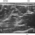
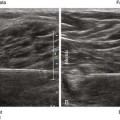
Twelve pairs of intercostal nerves lie within or near the inferior groove of each corresponding rib. These nerves supply the skin and chest wall skeletal muscles. An intercostal artery and vein accompany each nerve and lie superior to it. Intercostal nerves are difficult to image with ultrasound because they are small and often covered by the caudal edge of the corresponding rib.1 Proximal intercostal nerves are found in the classic subcostal position in 17%, in the midzone in 73%, and in the inferior supracostal position in 10% of anatomic specimens. The intercostal nerves migrate away from the ribs near the midaxillary line. Doppler ultrasound has been used to locate intercostal arteries for intercostal block.2 Intercostal arteries are 3 to 4 mm in diameter and can be detected in an acoustic window 4 cm from the midline.3 Doppler measurements of the intercostal arteries are possible from T4 and lower. Ultrasound-guided intercostal nerve block has been used for acute and chronic pain management.4 Intercostal nerve blocks can be used for breast surgery and are best placed at T3, T4, and T5 for this procedure. Another common application is for thoracic trauma and chest tube placement.5