• Gastrointestinal tract: Most common internal organ system involvement (80-90%)
Esophagus > duodenum > anorectal > small bowel > colon
• Small bowel
Marked dilatation of small bowel, especially duodenum and jejunum
Duodenal findings identical to SMA syndrome
“Hidebound” small bowel: Atonic with closely spaced thin folds, sacculations (pathognomonic of scleroderma)
Prolonged transit time with barium retention in duodenum and small bowel up to 24 hours
± pneumatosis intestinalis and pneumoperitoneum
± transient, nonobstructive intussusceptions
• Colon
Sacculations on border of transverse and descending colon
Loss of haustrations
Stercoral ulceration (from retained fecal material in rectosigmoid)
TOP DIFFERENTIAL DIAGNOSES
• SMA syndrome
• Celiac-sprue disease
• Ileus
DIAGNOSTIC CHECKLIST
• Markedly dilated atonic small bowel with thin, crowded circular folds and delayed barium transit time
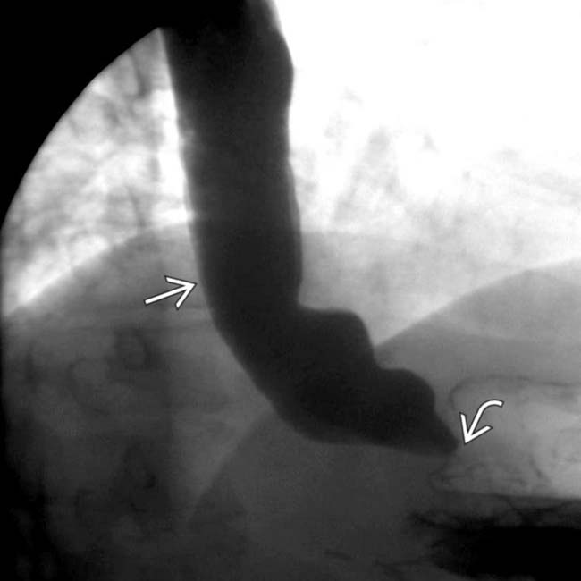
(Left) This 50-year-old man has diffuse scleroderma with progressive dysphagia & abdominal bloating. A film from the upper GI small bowel follow-through (SBFT) shows a dilated, atonic esophagus that is slow to empty due to a distal esophageal, peptic stricture .
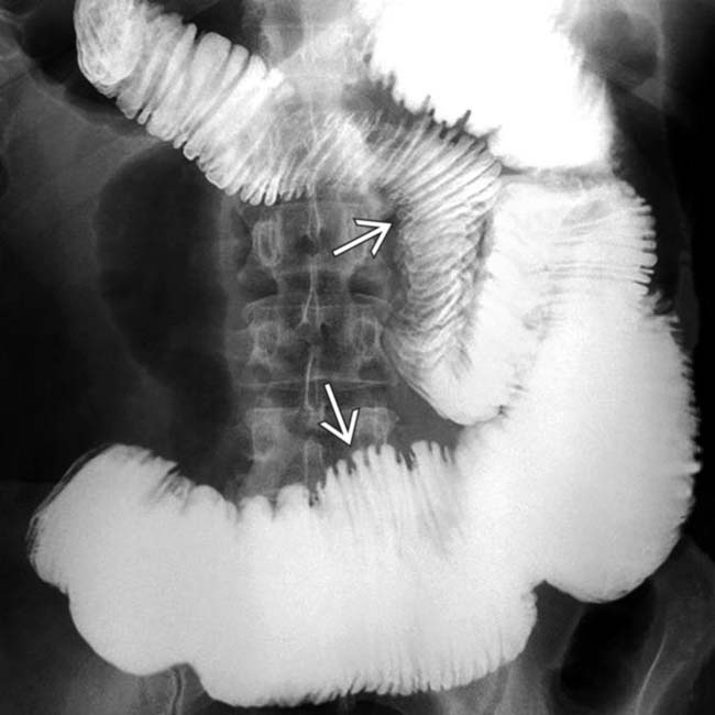
(Right) A 90-minute film (same case & study) from SBFT shows classic scleroderma of the small bowel with dilated, atonic jejunum & closely spaced, thin transverse folds with slow transit. “Pseudo-obstruction” is another descriptive term relevant to this case.
(Left) Axial CECT in a 40-year-old woman demonstrates closely packed, thin small bowel folds and diffusely dilated lumen, classic features of scleroderma with pseudo-obstruction.
(Right) Coronal CECT in the same patient demonstrates the dilated small bowel with a “hidebound” appearance of closely packed, thin folds (particularly in the jejunum), a characteristic feature of scleroderma. Also note the disproportionate dilation of the duodenum , another common feature of scleroderma.
TERMINOLOGY
Synonyms
• Progressive systemic sclerosis
Definitions
• Multisystem disorder of small vessels and connective tissue of unknown etiology
IMAGING
General Features
• Best diagnostic clue
Dilated, atonic small bowel with crowded folds and wide-mouthed sacculations
• Other general features
Multisystemic disorder with immunologic and inflammatory changes
Characterized by atrophy, fibrosis, sclerosis of skin, vessels, and organs
Involves skin and parenchyma of multiple organs
– GI tract, lungs, heart, kidneys, and nervous system
Gastrointestinal tract (GI) scleroderma
– 2nd most common manifestation after skin changes (80-90% of patients)
– Most common sites: Esophagus > duodenum > anorectal > small bowel > colon
– Most frequent cause of chronic intestinal pseudo-obstruction
Scleroderma classified into 2 types
– Diffuse scleroderma
– CREST syndrome (more benign course)
Diffuse scleroderma: Cutaneous and visceral involvement
– Severe interstitial pulmonary fibrosis
– Organ failure more likely
– Associated with antitopoisomerase I antibody (anti-Scl 70)
CREST syndrome: Less cutaneous and visceral involvement
– C alcinosis of skin
– R aynaud phenomenon
– E sophageal dysmotility
– S clerodactyly
– T elangiectasia
Radiographic Findings
• Esophagram
Atony, aperistalsis: Lower 2/3 of esophagus (smooth muscle)
Mild to moderate dilation of esophagus
Patulous lower esophageal sphincter: Early finding

 “Hidebound” small bowel: Atonic with closely spaced thin folds, sacculations (pathognomonic of scleroderma)


 that is slow to empty due to a distal esophageal, peptic stricture
  .
.
 with slow transit. “Pseudo-obstruction” is another descriptive term relevant to this case.
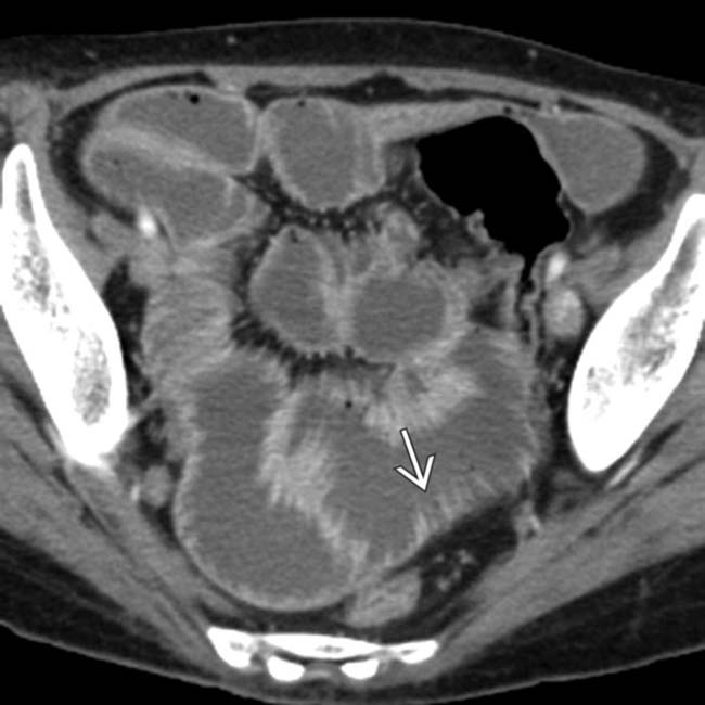
 and diffusely dilated lumen, classic features of scleroderma with pseudo-obstruction.
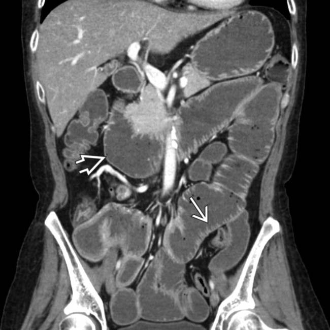
 (particularly in the jejunum), a characteristic feature of scleroderma. Also note the disproportionate dilation of the duodenum
  , another common feature of scleroderma.


 Marked dilatation of small bowel (particularly 2nd and 3rd parts of duodenum and jejunum)
 Pathognomonic: Hidebound sign of small bowel


