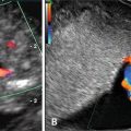
Fig. 20.1
Approaches to CVS: transcervical (red horizontal arrow) and transabdominal (green vertical arrow)
In our own experience, we perform the TC approach in about 70 % of singletons. Less experienced operators have a much higher proportion of TA cases because TA is usually easier for the inexperienced physician, as it is very comparable to doing an amniocentesis. The TC approach is more difficult and requires more experience to be competent and safe. Both approaches require a three-dimensional (3D) appreciation of the anatomy to be interpreted from 2D ultrasound. We have found that there is a divide that cannot be overcome just by experience between those physicians and sonographers who are capable of thinking and acting in 3D and those who cannot.
We have also seen operators who do not perform TC procedures have use contortions of lifting the uterus vaginally to make it reachable abdominally. We think this is inappropriate and increases the risk of procedures when they are done by the wrong approach for the given location. We believe that a CVS operator must be proficient in both approaches.
Other factors must be considered before attempting CVS. At times the patient gives a history of genital herpes simplex or a recent group B streptococcus (GBS) infection. Such cases should be individualized, and the small or theoretical risk of introducing an infection into the fetal-placental tissues should be discussed with the patient. TA-CVS or amniocentesis are usually offered when a significant risk of active GBS is present, as data are in favor that uterine infection, in such cases, might occur almost entirely after TC aspiration [21]. The choice of whether to perform TA-CVS or TC-CVS should be made according to the experienced operator’s judgment, based on the previously described conditions and in accordance with the patient’s bacterial/fungal cervical carrier status when known [14].
Safety
Over the past three decades, multiple reports from individual centers have demonstrated the safety and low rates of pregnancy loss following CVS [22, 23]. There is a large amount of literature, which will not be extensively reviewed here, from the 1980s and 1990s that has detailed the development of CVS as a clinical procedure and led to the US Food and Drug Administration’s removal of the restrictions on the use of the CVS catheter in 1989. The large majority of these projects did not find any increased risk of CVS over amniocentesis, but overall loss rates of both procedures were considerably higher than claimed by many operators of both procedures [22, 23].
Three studies in the past decade have been very important in our current understanding. The first was a meta-analysis published in 2007 by Mujezinovic and Alfirevic [5]. Because of the higher background rate of loss in the first trimester, CVS would be expected to have a higher overall loss rate. Yet it is impossible precisely to parse which losses are procedure-related, as opposed to background. What they reported is that, in experienced centers, the loss rates within 2 weeks of the procedure, up to 24 weeks and at term were essentially identical (Table 20.1).
Table 20.1
Amniocentesis and CVS loss ratesa
Loss before | Amniocentesis (%) | CVS (%) |
|---|---|---|
14 days | 0.6 | 0.7 |
24 weeks gestation | 0.9 | 1.0 |
Total | 1.9 | 2.0 |
The second study is the recent ongoing experience of the Danish first-trimester screening program, which over the past several years has maintained a very high detection rate for Down syndrome (>90 %) while cutting their false positive rates in half. As a consequence of first-trimester screening, the utilization of CVS in Denmark has been three times that of amniocentesis. Their data show that the procedure risk of CVS is as low (and possibly lower) than amniocentesis [24]. Furthermore, the incidence of late complications such as late demise was significantly lower in CVS patients than amniocentesis patients.
Recently Akolekar published a meta-analysis that demonstrated that overall procedure loss rates for CVS and amniocentesis were much lower than previously shown, and not different from each other—approximately 1/500 [6]. The primary confusion is the difference in background loss rates, which actually explain the vast majority of any differences between the two procedures.
Overall, we counsel our patients as to a 1/400 procedural risk of either CVS or amniocentesis in very experienced hands. These risks may, in fact, err on the side of being too high in our hands, but we do not want patients taking these decisions frivolously without considering the possibility of loss. Importantly, both procedures have considerably higher risks in inexperienced physicians or those who cannot visualize the anatomy in a 3D manner as described above. As such we believe that both CVS and amniocentesis should be performed preferably in “centers of excellence” by physicians, with experienced sonographers, who perform the procedure regularly—and not as an occasional item.
Complications of Chorionic Villus Sampling
Bleeding
Infection
Since the initial development of TC-CVS, there has been concern that TC-CVS would introduce vaginal flora into the uterus. This possibility was confirmed by cultures that isolated bacteria from up to 30 % of catheters used for CVS [26, 27]. In clinical practice, however, the incidence of post-CVS chorioamnionitis is low [9, 10]. Infection following TA-CVS also occurs and has been demonstrated, at least in some cases, to be secondary to bowel flora introduced by inadvertent puncture by the sampling needle.
Rupture of Membranes
Gross rupture of the membranes days to weeks after the procedure is acknowledged as a possible post-CVS complication. Rupture can result from either mechanical or chemical injury to the chorion, allowing exposure of the amnion to subsequent damage or infection. One group reported a 0.3 % incidence of delayed rupture of the membranes following CVS [28], a rate confirmed by Brambati et al. [29]. Unexplained, mid-trimester oligohydramnios has also been suggested as being a rare complication of TC-CVS.
Risk of Fetal Abnormalities Following Chorionic Villus Sampling
In the early 1990s it was suggested that CVS may be associated with specific fetal malformations, particularly limb reduction defects (LRDs). Today, based on the published data, it appears safe to state that there is no increased risk for LRDs, or any other birth defect, when CVS is performed at >70 days of gestation [30–33].
The first suggestion of an increased risk for fetal abnormalities following CVS was reported by Firth et al. [7]. In a series of 539 CVS-exposed pregnancies, there were five infants with severe limb abnormalities in a cohort of 289 pregnancies sampled by TA-CVS at 55–66 days gestation. Four of these infants had the unusual and very rare oromandibular-limb hypogenesis (OLH) syndrome, and the fifth had a terminal transverse LRD. OLH syndrome occurs in 1 per 175,000 live births [34], and LRDs normally occur in 1 per 1690 births [35]. Thus, the occurrence of these abnormalities in more than 1 % of CVS-sampled cases raised a high level of suspicion. Subsequently, other groups reported the occurrence of LRDs and OLH following “early” CVS [36–41]. In 1992, a case–control study using the Italian Multi-Center Birth Defects Registry, reported an odds ratio of 11.3 (95 % CI 5.6–21.3) for transverse limb abnormalities following first-trimester CVS [36]. However, when stratified by gestational age at sampling, pregnancies sampled prior to 70 days had a 19.7 % increased risk of transverse limb reduction defects, while patients sampled later did not demonstrate significantly increased risk. Other case–control studies, however, have not seen any association of CVS with LRDs [31, 32].
The risk for fetal malformations is real when CVS is done at an earlier gestation age, i.e., during the period of limb genesis at 6–7 weeks [7, 8, 38]. Brambati et al., practicing in Milan, have a large population at risk for β thalassemia. The population is also predominantly Catholic, for whom abortion at any gestational age is religiously proscribed. However, to assuage the guilt of wanting diagnosis, they wanted it done as early as technically possible. They reported a 1.6 % incidence of severe LRDs in a group of patients sampled at 6 and 7 weeks gestation [38]. This rate decreased to 0.1 % at 8–9 weeks. These data support the assumption that early gestational sampling and excessive placental trauma may be etiologic in the reported clusters of post-CVS LRDs.
In the USA, the population group most interested in early CVS has been the Orthodox Jewish community—at high risk for Tay Sachs and other Ashkenazi diseases. In the observant Jewish community, abortion is permitted, by religion, until 40 days post-conception (7 weeks 5 days from LMP). Wapner and Evans have shown that, in very experienced centers, CVS can be safely and reliably performed, even in very early gestation [30]. In a study they conducted, CVS was performed at less than 8 weeks gestation in a population of Orthodox Jews. Of the 82 cases of early CVS, there was only a single case of severe LRDs, a rate of 1.6 %. While this risk is considerably higher than when done at the usual time, we believe that in comparison to a 25 % risk of lethal disorder and with proper genetic counseling, it could be a reasonable decision for a couple to make. However, in the one case, despite the patient having had three previous early CVSs, they chose to sue the doctor stating that they did not “know.” As a result of the legal exposure, virtually all centers that were willing to do the procedure, stopped performing it.
The question whether CVS sampling after 10 weeks has the potential of causing more subtle defects, such as shortening of the distal phalanx or nail hypoplasia, was a major concern, debated thoroughly in the literature [40, 42]. The overall incidence of LRDs after CVS is estimated to be 1 in 1881 (ranging from 5.2 to 5.7 per 10,000), compared with 1 in 1642 (ranging from 4.8 to 5.97 per 10,000) in the general population [33, 42]; hence, there are no data to substantiate this concern. As noted, in most experienced centers performing CVS after 10 weeks, no increase in limb defects of any type was observed [31, 33, 41, 42].
Perinatal Complications
No increases in preterm labor, premature rupture of the membranes, small-for-gestational age infants, maternal morbidity, or other obstetric complications have occurred in sampled patients [43]. Although the Canadian Collaborative Study showed an increased prenatal mortality in CVS sampled patients, with the greatest imbalance being beyond 28 weeks, no obvious recurrent event was identified [10]. To date, CVS is not considered to harbor additional prenatal complication as long as the procedure is performed by an experienced operator and after 10 weeks gestation.
Long-Term Infant Development
Long-term infant follow-up has been performed by Chinese investigators who evaluated 53 children from their initial placental biopsy experience of the 1970s. All were reported in good health, with normal development and school performance [44]. Schaap et al. [45] obtained long-term follow-up data after CVS and amniocentesis and found no significant differences for neonatal and pediatric morbidity. Based on their data, the authors concluded that TC-CVS performed around 10 weeks gestation is not associated with an increased frequency of congenital malformations, compared with second trimester amniocentesis.
Accuracy of CVS Cytogenetic Results
A major concern with all prenatal diagnostic procedures is the possibility of discordance between the prenatal cytogenetic diagnosis and the actual fetal karyotype. With CVS, these discrepancies can occur from either maternal tissue contamination or from true biologic differences between the extraembryonic tissue (i.e., placenta) and the fetus. Fortunately, genetic evaluation of chorionic villi provides a high degree of success and accuracy, particularly in regard to the diagnosis of common trisomies [46]. In the late 1980s, the US Collaborative Study revealed a 99.7 % rate of successful cytogenetic diagnosis, with 1.1 % of the patients requiring a second diagnostic test, such as amniocentesis or fetal blood analysis, to further interpret the results [46]. In our own experience, a follow-up amniocentesis is needed about 0.5 % of the time.
Clinical errors or misinterpretation are rare, however, and the need for repeat testing continues to decrease, as more knowledge about the characteristics of chorionic villi is obtained. Indeed, studies [23, 47] have demonstrated that CVS is associated with a low rate of maternal cell contamination or chromosomal abnormalities confined to the placenta, as will be described below. For example, tetraploidy on CVS FISH and culture is seen in about 0.5 % of cases and is known to almost always be associated with a normal diploid fetus.
Maternal Cell Contamination (MMC)
Contamination of samples with a significant amount of maternal decidual tissue may lead to diagnostic errors, underlining the importance of preventing this occurrence [27, 47]. Generally, decidual contamination in CVS is almost always due to a small sample size, making appropriate tissue selection difficult. In experienced centers, in which adequate quantities of tissue are available, this problem is rare, with clinically significant MCC occurring in less than 0.5 % of CVS procedures. It is standard to separate maternal tissue from the sample. The chorionic “fronds” are distinguished from the maternal decidua under the microscope, making decidual removal by careful dissection possible.
In recent years there has been much progress in the molecular techniques suitable for detection of MMC, allowing more accurate results in cases of molecular diagnoses, where MMC may jeopardize the validity of the test.
Confined Placental Mosaicism
True discrepancies between the karyotype of the villus and the actual fetal karyotype can occur, leading to either false-positive or false-negative clinical results. Although initially there was concern that this might invalidate CVS as a prenatal diagnostic tool, subsequent investigations have led not only to a clearer understanding of the clinical interpretation of villus tissue results, but also revealed new information about the etiology of pregnancy loss, possible causes of intrauterine growth restriction (IUGR), and the biologic mechanisms for uniparental disomy and associated clinical syndromes.
A chromosomal aberration that does not involve the fetal cell lineage will produce a confined placental mosaicism (CPM), in which the trophoblast and perhaps the extraembryonic mesoderm may demonstrate aneuploid cells, but the fetus is euploid. Several mechanisms may apply in pregnancies where CVS mosaicism or non-mosaic feto-placental discrepancies are detected.
Mosaicism occurs in about 0.5 % of all CVSs [48, 49] but is confirmed in the fetus in only 10–40 % of these cases. In contrast, amniocentesis mosaicism is observed in only 0.3 % of cultures but, when found, is confirmed in the fetus in ~70 % of cases [50, 51]. These feto-placental discrepancies are known to occur because the chorionic villi consist of a combination of extra embryonic tissue of different sources that become separated and distinct from those of the embryo in early developmental stages. Specifically, at the 32–64-celled blastocyst, only 3–4 blastomeres differentiate into the inner cell mass (ICM), which forms the embryo, mesenchymal core of the chorionic villi, the amnion, yolk sac, and chorion, whereas the rest of the cells become the precursors of the extraembryonic tissues [52].
The probability of mosaic or non-mosaic trisomy in the fetus itself depends on the placental lineages in which the trisomic cell line was found. CVS culture represents the villous mesenchymal core and therefore reflects the chromosomal constitution of the fetus proper to a greater extent than the direct preparation, which represent the chorionic ectoderm, farther removed from the fetus. Thus, if a mosaic chromosomal aberration is detected on both direct preparation and long-term culture, it is more likely to represent a true mosaicism of the fetus [49]. Nevertheless, in gestations involving mosaic trisomic villous mesenchyme (with or without evidence of trisomy in direct cytotrophoblast examination), it is our usual policy to further examine the fetal karyotype by amniocentesis and perform a thorough fetal ultrasound scan in order to rule out fetal malformations.
Uniparental disomy (UPD) is another adverse outcome that may be associated with CPM. In UPD, both chromosome of a given pair are inherited from a single parent, rather than one from each. UPD results when the original trisomic embryo is “rescued” by the loss of the one extra chromosome. Because in the trisomic embryos two of chromosomes come from one parent and one from the other, there is a theoretical 1 in 3 chance that the two remaining chromosomes originate from the same parent, leading to UPD. This may have clinical consequences if the chromosome involved harbors imprinted genes whose expression vary according to the parent of origin or if the two remaining chromosomes carry a mutant recessive gene, creating a homozygous state. In general, UPD has been reported for almost every chromosomal pair, although clinical consequences have been observed mainly in cases involving specific chromosomes (i.e., chromosomes 2, 6, 7, 10, 11, 14, 15, 16, 20) and depending on the parent of origin [53]. For instance, despite a relative high frequency of CPM for trisomy 2 and trisomy 7, maternal UPD (2) and maternal UPD (7) have only been reported rarely [54, 55].
A significant CPM involves chromosome 15 and is encountered in 27/100,000 samples [56]. This is associated with risk for UPD (15) which may lead to well-recognized clinical syndromes. Chromosome 15 is known to carry genes that are subject to both paternal and maternal imprinting. Maternal UPD (15), resulting from the relatively more common maternally derived trisomy 15, causes the Prader–Willi syndrome. In contrast, paternal UPD (15) caused by rescue of the less common paternal trisomy 15, results in the less frequent Angelman syndrome.
In rare cases, CPM for trisomy 15 offers the important clue that UPD may be present in the “chromosomally normal” fetus, which may be at risk of having Prader–Willi/Angelman syndrome [57, 58]. For this reason, cases in which CVS reveals trisomy 15 (either complete or mosaic) should be evaluated for UPD if the amniotic fluid demonstrates an apparently euploid fetus [56].
Early Amniocentesis
Early amniocentesis is a procedure that has come and gone. It is a first-trimester procedure, i.e., performed before 14 weeks of gestation (usually from 11 + 0 to 13 + 6) [59, 60]. Some series have included procedures as early as 9 + 0 weeks. Traditional amniocentesis is usually performed after 15 + 0 weeks of gestation; invasive procedures between 14 + 0 and 14 + 6 are usually considered early and have been included in some series but not others [61]. Since 1987, various sized observational studies on EA reported rates of procedure-related fetal loss from 1.4 to 8.1 % (see Table 20.1). Early series concluded that EA is an appropriate technique for early diagnosis but is associated with an increased fetal loss rate [62, 63]. Assel et al. [64] compared EA with mid-trimester amniocentesis and found a significant increased post procedure fetal loss rate (1.8 % vs. 0.4 %) [65].
A prospective partially randomized study by Nicolaides et al. [66, 67] showed a higher rate of fetal loss after EA compared with CVS (4.9 % vs. 2.1 %), which was significant for pregnancies at 10–11 weeks but not significant for the 12–13 weeks gestation period.
The CEMAT study compared EA between 11 + 0 and 12 + 6 weeks with standard amniocentesis (15 + 0–16 + 6). This multicenter randomized trial reporting on 1916 EA procedures showed an increased total pregnancy loss (pre-procedure and post procedure losses including intrauterine and neonatal deaths) with the EA procedure (7.6 % vs. 5.9 %; P = 0.012) [68].
Post-procedure Amniotic Fluid Leakage
Besides fetal loss, additional complications have been reported as being directly related to EA procedures. Leakage of amniotic fluid after EA is concerning because of the risk for infection, miscarriage, preterm labor/delivery, and fetal neonatal complications. The reported incidence varies from 0 to 4.6 %. For example, the CEMAT study reported an increased rate of fluid leakage that was statistically significant before 22 weeks of gestation, when EA were compared with standard amniocentesis (3.5 % vs. 1.7 %) [68]. Many reports, however, have associated EA and congenital abnormalities. The lower limb extremities have increased susceptibility with temporary disturbances from a diminution in intra-amniotic volume [69]. Second trimester procedures do not have any such association [70]. However, the CEMAT [68] trial and several others showed a significant increased rate of a foot anomaly (1.3 % vs. 0.1 %; P = 001) for EA from 11 + 0 to 12 + 0 week. Tharmaratnam et al. [71] reported a rate of fixed flexion deformities of 1.6 %. They showed a positive association with the amount of amniotic fluid removed and the rate of musculoskeletal deformities [71].
An international randomized trial of late first-trimester invasive prenatal diagnosis to assess the safety and accuracy of amniocentesis and TA-CVS performed at 11–14 weeks was reported [47]. A fourfold increase in the rate of talipes equinovarus was observed in cases where early amniocentesis was the technique used. The authors concluded that amniocentesis at, or before, 13 weeks carries an increased risk for this specific limb defect and an additional increase in early, unintended pregnancy loss. In another study, Alfirevic et al. [72] have analyzed 14 randomized studies from the Cochrane Pregnancy and Childbirth Group Trials Registry and from the Cochrane Central Registry and Control Trials, in order to assess the safety and accuracy of the various invasive procedures employed for early prenatal diagnosis. Based on their results, they concluded that early amniocentesis is not a safe alternative to second trimester amniocentesis because of increased pregnancy loss (relative risk 1.29), and higher rates of talipes equinovarus (relative risk 6.43). Early amniocentesis has been essentially completely abandoned as CVS is a clearly safer procedure in the first trimester and, arguably, at 14 weeks as well.
In conclusion, for first-trimester diagnosis, either TA-CVS or TC-CVS are the clinically appropriate methods. We believe utilization of both CVS methods is necessary to have the most complete, practical, and safe approach to first-trimester diagnosis. EA carries a significant risk for fetal loss and fetal malformations. We have not found a situation in which EA was the appropriate method for a patient in about 20 years.
Fetal Reduction
Fetal reduction (FR) has changed considerably over the last 25 years since we first published on the subject [15, 73]. These changes have taken place in medical technology outcomes, patient choices, and the larger demographic and cultural shifts that are driving the pace and direction of change.
At its core, FR started out as a way of managing pregnancies in which the risks to both mother and fetuses from carrying multiple embryos were extreme. Selective termination (as it was called then) of some of the embryos to increase the viability of the remaining ones and reduce the risk of morbidity and mortality for the mother was a desperate approach to salvage the situation. As with numerous other technological changes, what began as a dominant concern with matters of life and death has eventually become accepted. Then indications transform from crisis “life and death” into issues of quality of life [74, 75].
FR was developed as a clinical procedure in the 1980s, when a small number of clinicians in both the USA and Europe attempted to reduce the usual and high adverse sequelae of multifetal pregnancies, by selectively terminating or reducing the number of fetuses to a more manageable number. The first European reports by Dumez and Oury [20], and the first American report by Evans et al. [73], followed by a further report by Berkowitz et al. [76], and later Wapner et al. [77] described a surgical approach to improve the outcome in such cases.
Multiple papers in the 1990s demonstrated that with triplets or more, there was clear improvement in reducing to twins. Numerous papers argued whether triplets had better outcomes “reduced” or not. Yaron et al. [78] compared triplets-to-twins data to unreduced triplets with two large cohorts of twins. The data showed substantial improvement of reduced twins as compared to triplets. The data from the 2001 collaborative series and others suggested that pregnancy outcomes for cases starting at triplets or even quadruplets reduced to twins at about 12 weeks do fundamentally as well as starting as twins.
Overall, statistics on reductions have improved noticeably over these past 25 years [14, 79, 80]. In the early 1990s, when half the cases were quadruplets or more, loss rates (up to 24 weeks) were 13 %. Early premature deliveries were an additional 10 %. Now, overall with decreasing starting numbers, better ultrasound, better understanding of zygosity, and a limited number of practitioners with extensive experience accounting for a high percentage of reductions, losses are overall down to about 4 %. Counseling must be tailored to specific starting and finishing numbers and the experience of the operator.
Related posts:
 Aneuploidy Screening: The Ongoing Role of First-Trimester Ultrasound
 Three-Dimensional Ultrasound: A Role in Early Pregnancy?
 The Fetal Heart in Early Pregnancy
 First-Trimester Ultrasound in Gestational Trophoblastic Disease
 A Consequence of Cesarean Delivery: First-Trimester Cesarean Scar Pregnancy
 Fetal Anomalies
Stay updated, free articles. Join our Telegram channel

Full access? Get Clinical Tree


