Acute ischemia: Clinical triad of sudden-onset abdominal pain, diarrhea, and vomiting
• Surgical treatment: Exploratory laparotomy, bowel resection, and mesenteric bypass to reestablish blood flow
DIAGNOSTIC CHECKLIST
• Image interpretation pearls
• Imaging findings vary due to many factors (e.g., acute vs. chronic; arterial vs. venous)
• Mesenteric venous occlusion causes more impressive wall thickening, mesenteric infiltration, and ascites than arterial occlusion
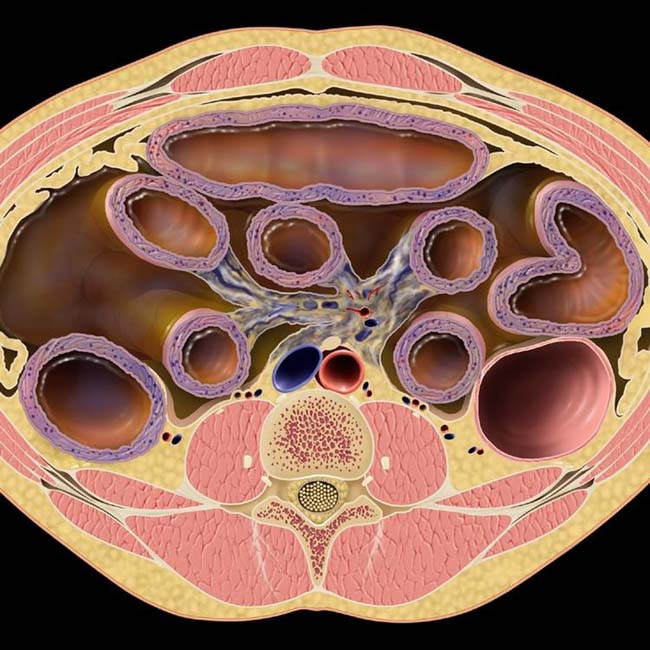
(Left) Graphic shows a dilated small bowel (SB) with thickened wall, ascites, and edematous mesentery, all findings seen with occlusion of the superior mesenteric vein.
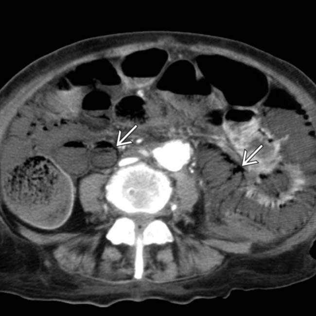
(Right) Axial CECT in an elderly woman with abdominal pain demonstrates dilated small bowel with pneumatosis . Portal venous gas was also present on other sections (not shown). Infarcted bowel was confirmed at surgery, and the patient died.
(Left) Oblique film from small bowel follow-through reveals luminal narrowing and a loss of normal small bowel fold pattern in the ileum , as well as dilation of the more proximal segments .
(Right) Superior mesenteric angiogram in the same patient illustrates a cutoff of some of the ileal branches of the superior mesenteric artery (SMA) with a lack of blood flow to the ileum. This appearance suggests an embolic source, as thrombosis would tend to involve the origin of the SMA.
TERMINOLOGY
Synonyms
• Acute mesenteric ischemia
Definitions
• Mesenteric arterial or venous narrowing or occlusion, leading to inadequate supply of nutrients and oxygen to small bowel (SB)
IMAGING
General Features
• Best diagnostic clue
Clot or narrowing of superior mesenteric artery (SMA) or superior mesenteric vein (SMV) with bowel wall thickening
Radiographic Findings
• Radiography
Multiple air-fluid levels; ileus pattern
Thickening of valvulae conniventes
Linear distribution of gas (pneumatosis intestinalis)
Fluoroscopic Findings
• Barium studies
Thickening of valvulae conniventes
“Thumbprinting” pattern: Intramural accumulation of blood distending submucosa → focally rounded mesenteric folds, especially along mesenteric border
“Stack of coins” pattern: Enlarged, smooth, straight, parallel folds perpendicular to longitudinal axis of SB (submucosal edema)
Strictures often seen with proximal bowel dilation
Mottled, frothy, bubbly, or linear collections of gas in bowel wall (pneumatosis intestinalis)
CT Findings
• CECT
Clot or reduced lumen in SMA, SMV, or other mesenteric vessels
Segmental thickening of bowel wall (> 3 mm)
Emboli usually observed at origin of SMA or 3-10 cm from SMA distal to middle colic artery
Lack of bowel mucosal enhancement due to compromised arterial flow
“Misty” mesentery: Mesenteric fat infiltrated by edema; more common with venous thrombosis
↑ attenuation of bowel wall due to submucosal hemorrhage or hyperemia


 . Portal venous gas was also present on other sections (not shown). Infarcted bowel was confirmed at surgery, and the patient died.
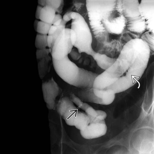
 , as well as dilation of the more proximal segments
  .
.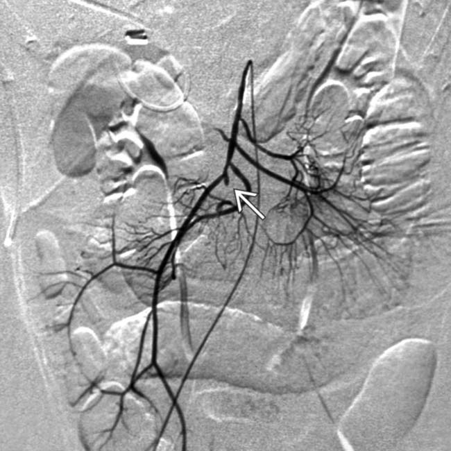
 of some of the ileal branches of the superior mesenteric artery (SMA) with a lack of blood flow to the ileum. This appearance suggests an embolic source, as thrombosis would tend to involve the origin of the SMA.


