Case presentation
A 4-year-old male presents with right forearm pain after falling while in a “bouncy house.” Apparently, the child was in the house with several other children and had a witnessed fall onto the arm. The parents noted that he had swelling to his right elbow. There is no other reported injury, and Emergency Medical Services applied a temporary splint and gave the child intranasal fentanyl (2 μg/kg) for pain management.
His physical examination is significant for right elbow pain and swelling. He has limited range of motion of the forearm at the elbow but he does not appear to have shoulder, upper arm, wrist, hand, or digit injury. He is grossly neurovascularly intact.
Imaging considerations
Plain radiography
Plain radiography is an appropriate imaging modality to utilize in patients with suspected skeletal injury. It is important to obtain adequate imaging, which includes anterior-posterior (AP) and lateral views of the entire forearm and elbow (important for suspected Monteggia fracture) and wrist (important for suspected Galeazzi fracture). ,
Computed tomography (CT)
CT does not generally play a role in the evaluation of patients with suspected Monteggia or Galeazzi fracture. CT may be indicated if injury at either the radiocapitellar or radioulnar joints is suspected but not confirmed on initial plain radiographs or if the injury is complex (e.g., comminuted fracture).
Magnetic resonance imaging (MRI)
This modality is generally not indicated in the initial evaluation of musculoskeletal injury. If there is a complex injury or neurologic symptoms, then MRI may be indicated; consultation with a pediatric orthopedic specialist would be advisable in these cases.
Imaging findings
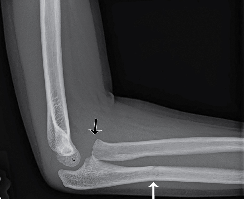
Plain radiographs of the patient’s right forearm, including AP and lateral views, were obtained and are provided here. There is a transverse fracture through the proximal to mid diaphysis of the right ulna, superimposed upon a bowing fracture of the ulnar diaphysis. There is an accompanying radial head dislocation ( Figs. 58.1 and 58.2 ).
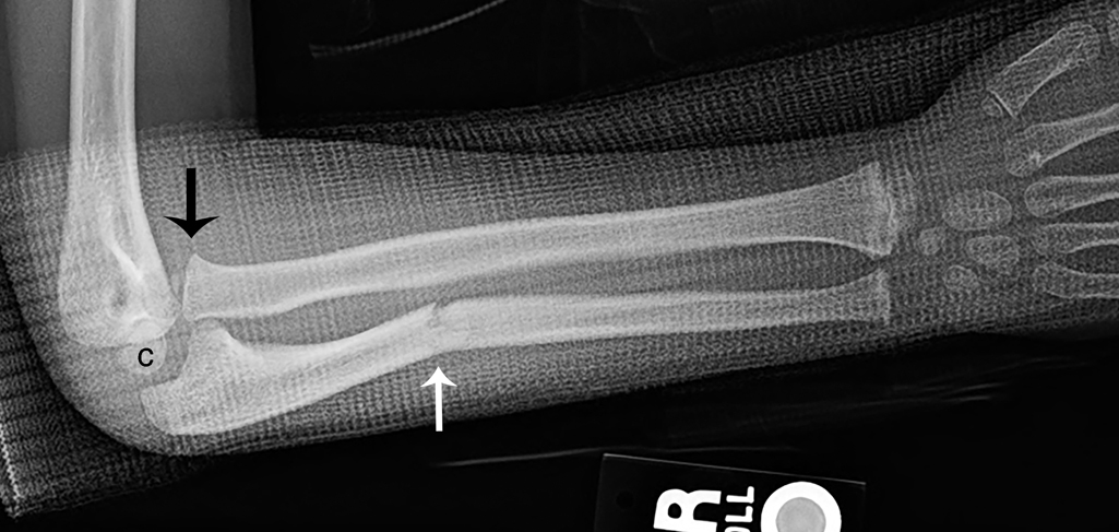
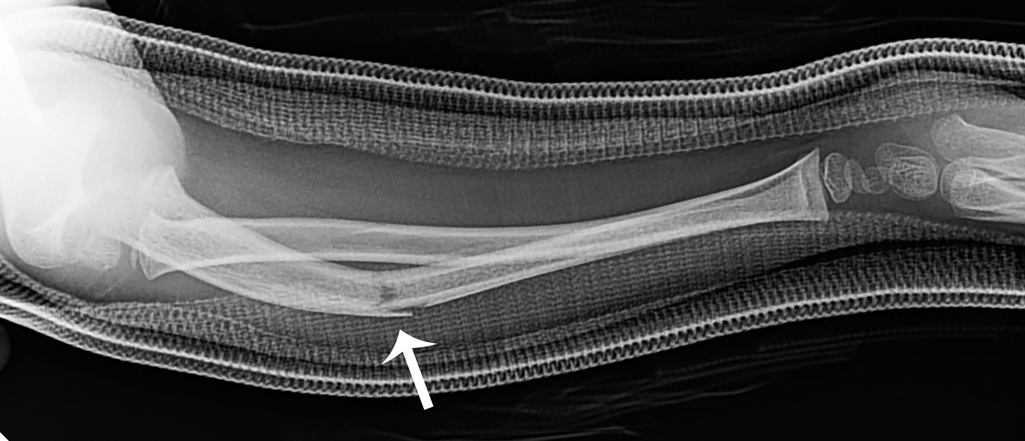
While the presence of the splint does obscure some detail, the process of removing the splint was challenging, leading to difficulties imaging the child. If a splint is applied prior to radiography, it should be removed if possible; if this is not able to be accomplished, then imaging should be performed with the best views that the clinical situation will allow.
For comparison, two additional cases are presented here:
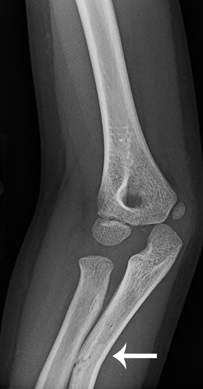
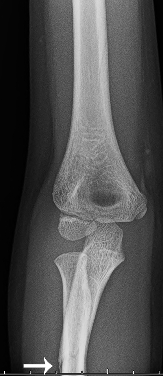
The child in Figs. 58.3–58.5 fell while riding her bicycle. These images demonstrate a nondisplaced fracture of the proximal third of the ulnar diaphysis, with anterior dislocation of the radial head, consistent with a Monteggia fracture. Note that the dislocation of the radial head is seen on the lateral view ( Fig. 58.3 ) and not adequately seen on the oblique and frontal views ( Figs. 58.4 and 58.5 ). This demonstrates the importance of obtaining a good-quality lateral view of the elbow region.