40.1 KUB control image
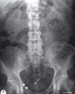
There are several small densities projected over the right side of the pelvis. Subsequent IVU images showed that two of these (arrowheads) were phleboliths (venous calcification) but there was also a vesico-ureteric junction (VUJ) calculus/stone (arrow)
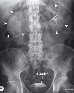
There is uptake of contrast by the renal parenchyma bilaterally forming an outline of both kidneys (arrowheads). On the left there is normal excretion of contrast into the renal collecting system (arrow). Contrast is also seen collecting in the bladder
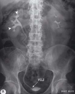
A ‘20 minute image’ demonstrated similar appearances with ‘clubbed’ or dilated calyces (arrowheads) and a column of contrast agent. Post-micturition this column persisted with the ureter filling from the VUJ upwards. The level of obstruction is identical to the position of the VUJ stone on the control image (see fig 40.1)
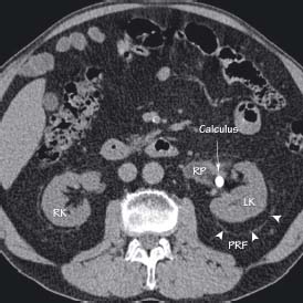
This patient had a history of recurrent renal colic due to renal calculi. Here a calculus is seen in the renal pelvis (RP) of the left kidney (LK). RK right kidney, PRF perirenal fascia
Intravenous urogram (IVU)
Related posts:


Stay updated, free articles. Join our Telegram channel

Full access? Get Clinical Tree


