Case presentation
A 4-year-old child presents after falling on an outstretched hand while running at school. He complains of pain to the right forearm and is splinted by the school nurse and given pain medicine by paramedics. There is no open abrasion or puncture to the skin. His vital signs are all age appropriate. There does not appear to be any injury to the head, neck, back, shoulder, upper arm, or wrist. He is grossly neurovascularly intact.
Imaging considerations
Most forearm fractures result from a fall onto an outstretched hand, and immediate pain control is key to completing a thorough examination as well as obtaining adequate radiographs. Intranasal fentanyl (2 mcg/kg, maximum single-dose 100 mcg) delivered via a mucosal atomizer device can achieve pain control within 10 minutes while radiographs and intravenous access are obtained.
Plain radiography
This is the imaging modality of choice when evaluating patients with suspected forearm fractures. A true anteroposterior view (AP) and lateral view of the forearm should include the wrist and distal humerus. A true lateral view of the forearm will illustrate partial overlapping of the radius and ulna at the proximal and distal ends and the elbow will be flexed at 90 degrees. If there is concern about an elbow fracture or dislocation, dedicated elbow radiographs should be obtained.
Ultrasound (US)
US has been shown to be a feasible method to diagnose isolated forearm fractures in resource-limited settings and has been used to guide reductions. The combination of US with clinical decision instruments has also been used in resource-rich settings to diagnose and manage treatment of distal forearm fractures with the intent to reduce radiograph use. Most studies have shown that time to train emergency physicians in diagnosing straightforward forearm fractures is short (i.e., 1 hour). The utility of US is more limited when evaluating midshaft and proximal forearm fractures that involve the elbow or more complicated injuries.
Advanced imaging (computed tomography and magnetic resonance imaging)
These imaging modalities are not indicated for the initial evaluation of forearm injuries. However, for complex fractures or if the extent of the fracture is not clear, these modalities may be utilized. Consultation with a Pediatric Orthopedic specialist is appropriate prior to employing these modalities.
Imaging findings
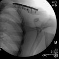
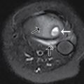
After appropriate pain medication administration, the patient had two-view imaging of the forearm. This demonstrated mid radial and ulnar diaphyseal fractures with mild volar angulation of the distal fracture fragments; the visualized portions of the elbow and distal humerus appear normal ( Figs. 57.1 and 57.2 ).


Case conclusion
Pediatric Orthopedics was consulted and the child was placed in a cast. Although the fracture did not require reduction, the fracture was molded during the cast application, and, due to good pain control, the patient tolerated this well. Follow-up was arranged. Imaging 2 months later demonstrated callous formation with near anatomic alignment ( Figs. 57.3 and 57.4 ).


Forearm fractures are the most common fractures in children, accounting for almost 50% of all childhood fractures. The radius and ulna are uniquely connected by an interosseous membrane longitudinally and by articulations at both the elbow and the wrist. If, by examination or radiograph, one bone is fractured, special attention should be paid to the other bone as well as proximal and distal joints for concurrent injuries. Physical examination should pay special attention for punctures to the skin with oozing blood, which may be a sign of an open fracture with a retracted segment. Sensory and motor examinations are important to assess for median, radial, and ulnar nerve neurapraxias. Forearm fractures can rarely have a complicating supracondylar fracture (also called a “floating elbow”), so providers should thoroughly examine the elbow and also evaluate for signs of compartment syndrome in these cases.
Distal forearm fractures should be evaluated by radiograph for physeal injuries and correlated for risk of Salter-Harris type I injuries by localized tenderness on examination. Buckle fractures (torus fracture) commonly occur at the distal metaphysis and are important to differentiate from nondisplaced greenstick fractures by radiograph. An ulnar styloid fracture is a distal avulsion fracture that often has a concurrent radial fracture, but avulsions at the base of the styloid itself may warrant surgical intervention.
Management of these injures depends on several factors, including the age of the child and severity of the fracture. Traditional management of displaced forearm fractures included closed reduction followed by immobilization with casting. , Fractures that are treated with closed reduction and immobilization do have a reported risk of radiographic loss of reduction of 5% to 75%; this risk is related to the amount of initial displacement and the inability to achieve anatomic alignment after reduction, among other factors. , Surgical management should be performed in patients with open fractures, fractures with neurovascular compromise, and fractures that have failed attempted closed reduction. , The most commonly utilized surgical technique is intramedullary fixation or internal fixation with plates/screws. ,
Special consideration should be given to bowing and greenstick fractures. Plastic deformities (or bowing fractures) will show bowing on radiograph without a clear fracture. Greenstick fractures are unique to pediatrics and will show a complete fracture on one side of the cortex and a buckling or bowing or the other side. Bowing fractures may be initially missed during radiographic interpretation. The management of these fractures varies, with some authors advocating that all bowing deformities should undergo reduction and others citing age and degree of deformity as criteria for reduction. , Greenstick fractures are generally treated with closed reduction and immobilization, although controversy exists as to whether the fracture should be completed and then reduced. Consultation with a pediatric orthopedic specialist is appropriate and helpful in determining management options in these patients.
For comparison, additional cases are presented here:
The patient in Figs. 57.5 and 57.6 fell off the monkey bars while playing at a park. Imaging of the forearm demonstrates plastic bowing fractures of the shafts of the radius and ulna, with apex posterior bowing. A nondisplaced greenstick fracture of the proximal shaft of the radius is also seen. Pediatric Orthopedics was consulted and spoke at length with the family, offering casting with close monitoring or reduction in the operating room. After the risks and benefits of each course of action were discussed, they family elected casting.


