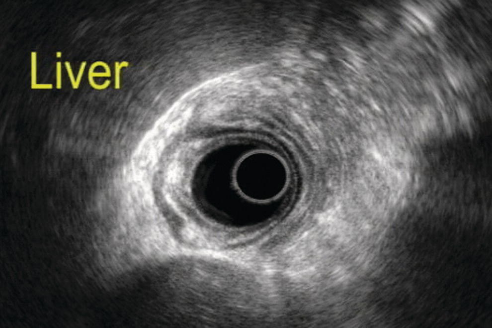
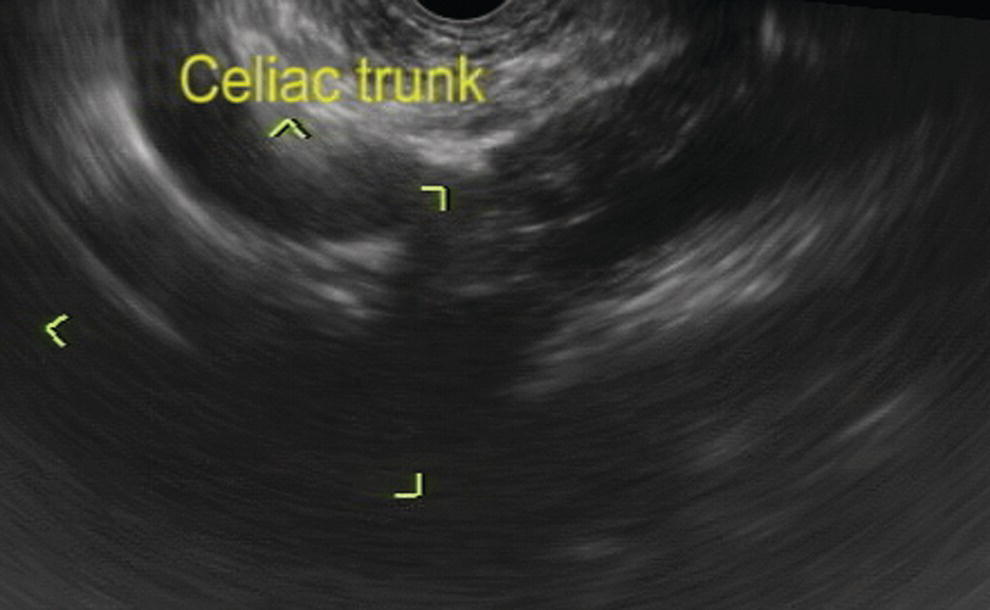
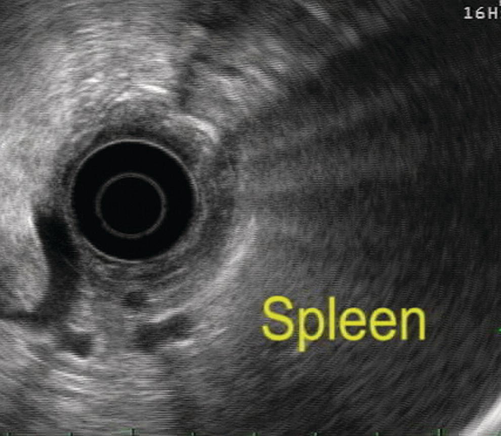
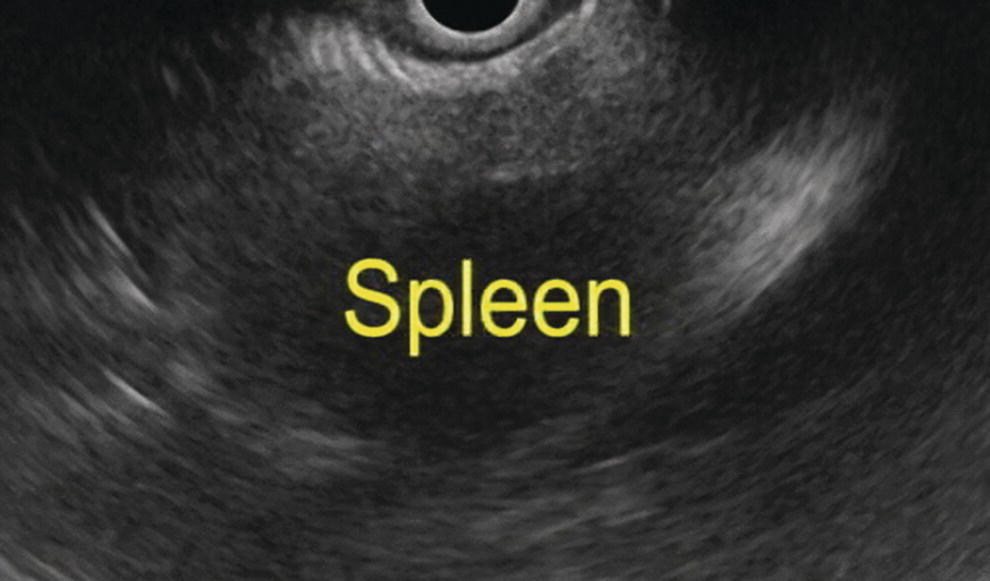
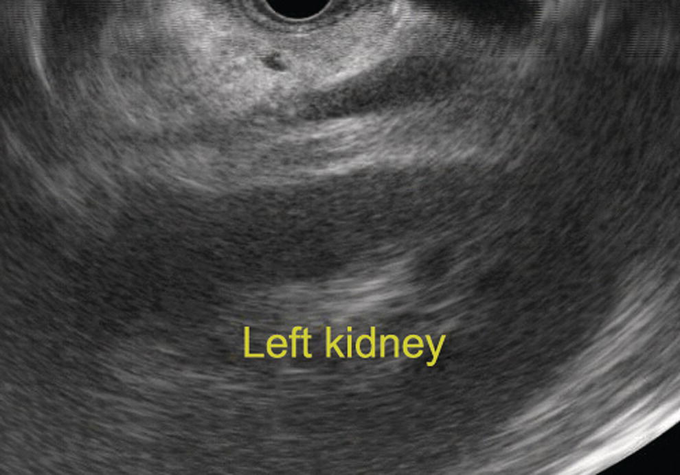
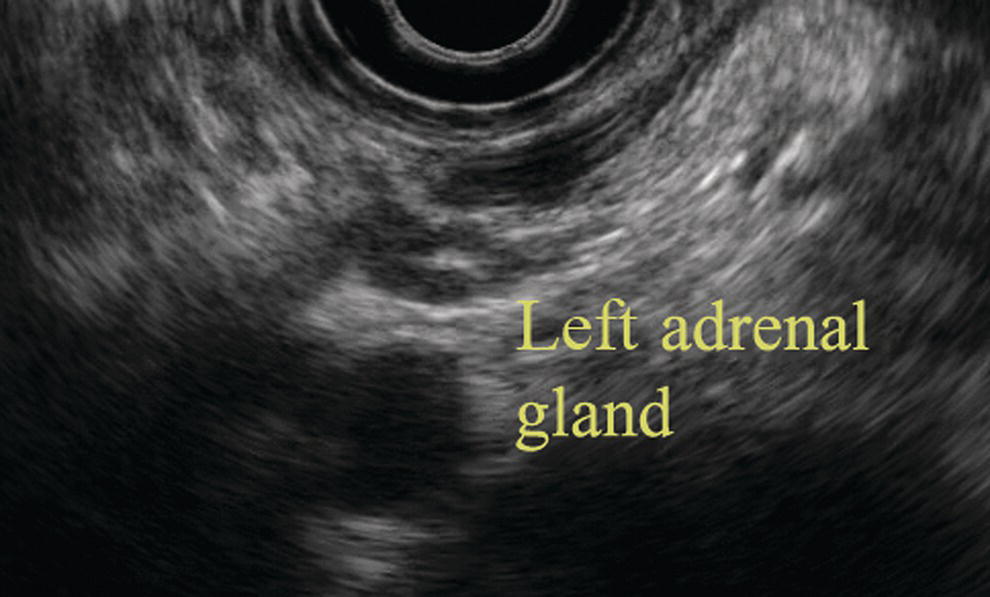
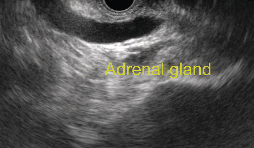
Nalini M. Guda1 and Marc F. Catalano2 1 University of Wisconsin, School of Medicine and Public Health, Pancreatobiliary Services, St. Luke’s Medical Center, Milwaukee, WI, USA 2 Medical College of Wisconsin, Pancreatobiliary Services, St. Luke’s Medical Center, Milwaukee, WI, USA This chapter describes the endosonographic features of the major organs of the abdomen: the liver, spleen, kidneys, and adrenal glands. Ultrasound features of the pancreas and bile duct are described elsewhere. The liver, spleen, kidneys, and adrenal glands (left side) are visualized from the stomach (Videos 7.1 and 7.2). As the radial probe is advanced through the esophagus into the gastric cardia, the liver is the predominant organ visualized. When positioning the abdominal aorta at the 6 o’clock position, the left lobe of the liver is seen anteriorly and medially to the right (Figure 7.1). The aorta, with a dark hypoechoic band which is the diaphragmatic crux, is seen immediately adjacent to the probe. In this position, near the hiatus, the hepatic veins are seen as anechoic structures, entering the inferior vena cava (IVC). In this position, possibly with left tip deflection, the spleen can be seen on the right of the screen. As the aorta is traced distally, maintaining its 6 o’clock position, the liver may still be seen anteriorly. Vascular structures can be differentiated from ductal structures by a thicker (echogenic) wall and the presence of flow. When advancing the echoprobe towards the antrum, the gallbladder is often visualized as an oval‐shaped anechoic structure. In this position, the porta hepatis can be seen with subtle tip deflection upwards. With the probe at the level of the diaphragmatic hiatus, the longitudinal aorta and celiac artery origin are the most recognizable reference points (Figure 7.2), demonstrated as tubular longitudinal structures. Here, rotation of the probe counterclockwise will bring into view the liver parenchyma and its vascular structures (Figure 7.3). Advancing the probe at the level of the pylorus and duodenal bulb, clockwise rotation and superior tip deflection brings into view the porta hepatis along with several vascular structures. Use of Doppler can differentiate arterial from venous structures as well as biliary structures. The entire liver is not visualized by endoscopic ultrasound (EUS). Despite this limitation, it is useful to carefully examine the liver since metastatic processes can be easily identified and biopsied and could lead to a change in clinical staging and management of a suspected tumor. The spleen appears as a homogeneous structure seen between the tail of the pancreas, left kidney, and gastric wall. With a radial scope it is imaged from the gastric cardia. It is similar to liver in echogenicity except that it is devoid of any ducts and vessels (Figure 7.4). It can be easier to follow the splenic vein after visualizing the pancreas from the gastroesophageal (GE) junction. The splenic artery, splenic vein, renal vein, and the left adrenal are usually visualized as well while attempting to scan the spleen. The splenic vein can be easily traced along the inferior aspect of the body and tail of the pancreas; however, the splenic artery is tortuous and it is difficult to follow its course to the celiac trunk. With a linear scope one has to scan inferior to the left kidney and laterally to visualize the spleen (Figure 7.5). Both the right and left kidneys can be visualized by EUS. The left kidney is readily identified from the fundus of the stomach. It has a complex echogenicity with hypoechoic parenchyma and hypertonic areas within representing the calyceal system. Figure 7.1 Image of the liver scanned by radial ultrasound. Figure 7.2 Celiac artery imaging by linear ultrasound. Figure 7.3 Image of liver by linear ultrasound. With the echoendoscope in the gastric cardia the left kidney is readily visualized. One can see the calyceal system and the renal vessels (Figure 7.6). To visualize the right kidney the echoscope is placed in the D‐3 position. Visualization of the IVC and aorta provide easily identifiable landmarks. Upon slow withdrawal, the image brings into view the right kidney immediately right of the probe along with the right renal vein and artery. As the kidney is traced proximally, lateral deflection of the tip may bring into view the right adrenal gland. Figure 7.4 Image of spleen by radial ultrasound. Figure 7.5 Image of spleen by linear ultrasound. With a linear scope one can withdraw the scope in the fundus posteriorly towards the pancreas until the tail is visualized and then push the scope inferiorly to see the left kidney (Figure 7.7). To view the right kidney, the probe is placed just below the level of the papilla and counterclockwise rotation brings into view the right kidney and its vascular structures. Slow withdrawal allows for visualization of the right adrenal glands. Figure 7.6 Image of left kidney by radial ultrasound. Figure 7.7 Image of left kidney by linear ultrasound. With the probe in the gastric fundus, the left adrenal gland can be found immediately proximal to the celiac artery and left of the aorta. It is seen as an echo‐poor, thin strip of tissue (“seagull” shaped). The left adrenal gland can also be located by tracing the left kidney cephalad, located medial to the upper pole of the kidney (Figure 7.8). Figure 7.8 Image of left adrenal gland by radial ultrasound. Figure 7.9 Image of left adrenal gland by linear ultrasound. With the probe at the level of the celiac trunk take‐off, slight clockwise rotation brings into view the left adrenal gland seen as a thin echo‐poor structure (Figure 7.9).
7
Liver, Spleen, and Kidneys: Radial and Linear
Introduction
Liver
Radial endosonography
Linear endosonography
Spleen
Kidney

Radial endosonography
Linear endosonography

Adrenal glands
Radial endosonography
Linear endosonography
Related posts:


Stay updated, free articles. Join our Telegram channel

Full access? Get Clinical Tree


