Figure 14.1
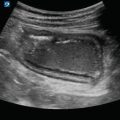
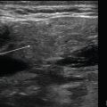
Comparison between vein and artery. Arteries (A) have a thick muscular wall that is pulsatile and maintains its circular structure with compression. Veins (V) have a thin wall with increased compliance and therefore lack a specific shape; they are easily compressible under pressure
Image Acquisition
Transducer Selection
Linear array.
Curvilinear transducer may be needed for obese patients.
Patient Position
For imaging the proximal lower extremity veins including common femoral, saphenous, superficial femoral, and deep femoral:
The patient should be placed in a supine position with their leg externally rotated, knee slightly flexed, and the head of the bed elevated or with the bed in reverse Trendelenburg.
Figure 14.2—Patient position for proximal veins.
Standard Exam Views
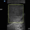
Place the transducer just inferior to the inguinal ligament on the anteromedial thigh to visualize the common femoral vein:
With the transducer perpendicular to the vein, apply direct and even downward pressure with the transducer and observe for complete compressibility of the vein:
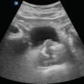
Figure 14.6—Common femoral vein compressed
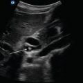
Slowly move the transducer distally tracing the common femoral vein until the area where the greater saphenous vein drains into the common femoral vein can be seen:
Figure 14.7—Common femoral vein with greater saphenous vein.
Video 14.3—Common femoral vein with greater saphenous vein.
Apply pressure and observe for collapse of the greater saphenous vein and the common femoral vein.
The greater saphenous vein is technically a superficial vein. However, it can easily travel into the common femoral vein and therefore will need anticoagulation [2].
Again, move the transducer distally until the bifurcation of the common femoral vein into the superficial and deep common femoral veins is visualized:
Trace the superficial femoral vein distally compressing every 2 cm until just proximal to the knee.
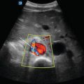
Next, place the transducer within the popliteal fossa and image the popliteal vein:
The popliteal vein will be located superficial or anterior to the popliteal artery.
Compress the popliteal vein and visualize complete occlusion of the vessel.
Figure 14.10—Popliteal vein.
Video 14.5—Popliteal vein.
Complete the scan by fanning the transducer distally to image the trifurcation of the popliteal vein into the anterior tibial, posterior tibial, and peroneal veins:
Apply pressure with the transducer and visualize compression of each vein.
Figure 14.11—Trifurcation.
Video 14.6—Trifurcation.

Figure 14.2
Patient position for proximal vein imaging. To assess the proximal lower extremity veins, externally rotate the patient’s leg and slightly flex the knee

Figure 14.3
Patient position for popliteal vein imaging. Flex the knee with slight external rotation of the patient’s leg

Figure 14.4


Transducer placement inferior to inguinal ligament. The transducer is placed just below the inguinal ligament perpendicular to the femoral vessels
Related posts:
Stay updated, free articles. Join our Telegram channel
Full access? Get Clinical Tree
