Lower Limb I: Pelvic Girdle, Sacrum, and Proximal Femur


Lower Limb I: Pelvic Girdle, Sacrum, and Proximal Femur


Pelvic Girdle

Fractures involving the structures of the pelvic girdle, which are usually sustained in motor vehicle accidents or falls from heights, represent only a small percentage of all skeletal injuries. Their importance, however, lies in the significant morbidity and mortality associated with them, which is usually caused by accompanying injury to the major blood vessels, nerves, and lower urinary tract. Because the clinical signs of pelvic trauma may not always be obvious, radiologic examination is essential to establish a correct diagnosis. Fractures of the acetabulum constitute approximately 20% of all pelvic fractures, and they may or may not be associated with dislocation in the hip joint. Fractures of the proximal (upper) femur, occasionally referred to as hip fractures, occur commonly in the elderly, often as a result of minimal injury. They are seen more frequently in women than in men (2:1), with intracapsular fractures of the proximal femur having an even higher female-to-male ratio (5:1).


Anatomic-Radiologic Considerations

The main imaging modalities used in the evaluation of traumatic conditions of the pelvic girdle, acetabulum, and proximal femur include conventional radiography and computed tomography (CT). Other ancillary techniques are also essential for a complete evaluation of concomitant soft-tissue and pelvic-organ injuries: angiography for the pelvic blood vessels and cystourethrography for the lower urinary tract. Radionuclide bone scan and magnetic resonance imaging (MRI) may also be necessary to disclose subtle fractures of the femoral neck and early stages of posttraumatic osteonecrosis of the femoral head.

The standard and special radiographic projections used to evaluate injury to the pelvic girdle and proximal femur include the anteroposterior view of the pelvis, the anterior and posterior oblique views of the pelvis, the anteroposterior view of the hip, and the frog-lateral view of the hip. At times, the groin-lateral or other special projections may also be required.

Most traumatic conditions involving the sacral wings, the iliac bones, the ischium, the pubis, and the femoral head and neck can be evaluated sufficiently on the anteroposterior projection of the pelvis and hip (Fig. 8.1). This view also demonstrates an important anatomic relation of the longitudinal axes of the femoral neck and shaft. Normally, the angle formed by these axes ranges from 125 to 135 degrees. This measurement is valuable in determining displacement in femoral neck fractures. A varus configuration is characterized by a decrease in this angle, and a valgus configuration by an increase in this angle (Fig. 8.2). The anteroposterior view, however, is frequently not sufficient to provide adequate evaluation of the entire sacral bone, the sacroiliac joints, and the acetabulum. Demonstration of the sacroiliac joints requires either a posteroanterior projection, which is obtained to greater advantage with 25 to 30 degrees caudal angulation of the radiographic tube, or an anteroposterior view with 30 to 35 degrees cephalad angulation. The latter projection, known as the Ferguson view, is also helpful in more effectively evaluating injury to the sacral bone and the pubic and ischial rami (Fig. 8.3). Oblique projections, known as Judet views, are necessary to evaluate the acetabulum. The anterior (internal) oblique projection helps delineate the iliopubic (anterior) column and the posterior lip (rim) of the acetabulum (Fig. 8.4). The posterior (external) oblique projection delineates the ilioischial (posterior) column and the anterior acetabular rim (Fig. 8.5). Of value in demonstrating the structures of the proximal femur and hip, the frog-lateral projection allows adequate evaluation of fractures of the femoral head and the greater and the lesser trochanters (Fig. 8.6). Demonstration of the anterior and posterior aspects of the femoral head as well as the anterior rim of the acetabulum may require a groin-lateral projection of the hip, which is particularly useful in evaluating anterior or posterior displacement of fragments in proximal femoral fractures and the degree of rotation of the femoral head. This projection, by providing an almost true lateral image of the proximal femur, also demonstrates an important anatomic feature, the angle of anteversion of the femoral neck, which normally ranges from 25 to 30 degrees (Fig. 8.7).

Ancillary imaging techniques play a crucial role in the evaluation of traumatic conditions of the pelvis and acetabulum, providing essential and often otherwise unobtainable information that helps the orthopedic surgeon determine the method of treatment and assess the prognosis of pelvic and acetabular fractures. Because the surgical management of such fractures is based on the stability of the fragments and the presence or absence of intraarticular extension of the fracture line and intraarticular fragments, CT examination is necessary to provide information that is not available from the standard and special projections of conventional radiography (Fig. 8.8; see also Figs. 8.22, 8.23, 8.24). In addition to ascertaining the size, number, and position of the major fragments and data about the condition of the weight-bearing parts of the joint and the configuration of the fracture fragments, CT can delineate soft tissue and concomitant injury to soft-tissue structures. However, in cases of severe injury when immediate surgical intervention is required, obtaining CT scans may be time-consuming and impractical. In such cases, conventional radiographs can be obtained more quickly, allowing more rapid recognition of the type of injury. CT is particularly effective in the postsurgical assessment of the alignment of fragments and fracture healing.


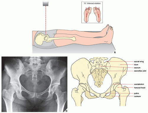
FIGURE 8.1 Anteroposterior view. (A) For the anteroposterior view of the pelvis and hip, the patient is supine with the feet in slight (15 degrees) internal rotation (inset), which compensates for the normal anteversion of the femoral neck (see Fig. 8.7B), elongating its image. For a view of the entire pelvis, the central beam is directed vertically toward the midportion of the pelvis; for selective examination of either hip joint, it is directed toward the affected femoral head. (B) The radiograph in this projection demonstrates the iliac bones, the sacrum, the pubis, and the ischium, as well as the femoral heads and necks and both the greater and the lesser trochanters. The acetabula are partially obscured by the overlying femoral heads, and the sacroiliac joints are seen en face.


FIGURE 8.2 Femoral shaft and neck angles. (A) The angle formed by the longitudinal axes of the femoral shaft and neck normally ranges from 125 to 135 degrees. In the evaluation of displacement in femoral neck fractures, a decrease in this angle (B) is known as a varus deformity, while an increase (C) characterizes a valgus deformity.


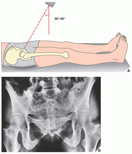
FIGURE 8.3 Ferguson view. (A) For the angled anteroposterior (Ferguson) view of the pelvis, the patient is in the same position as for the standard anteroposterior projection. The radiographic tube, however, is angled approximately 30 to 35 degrees cephalad, and the central beam is directed toward the midportion of the pelvis. (B) The radiograph in this projection provides a tangential view of the sacroiliac joints and the sacral bone. The pubic and ischial rami are also well demonstrated.


FIGURE 8.4 Anterior oblique view. (A) For the anterior oblique (Judet) view of the pelvis, the patient is supine and anteriorly rotated, with the affected hip elevated 45 degrees (inset). The central beam is directed vertically toward the affected hip. (B) On the radiograph in this projection, the iliopubic (anterior) column (arrows) (see Fig. 8.20) and the posterior lip (rim) of the acetabulum (open arrow) are well delineated.


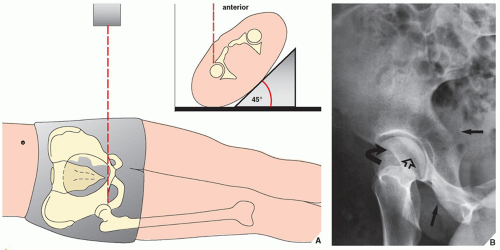
FIGURE 8.5 Posterior oblique view. (A) For the posterior oblique (Judet) view of the pelvis, the patient is supine and anteriorly rotated, with the unaffected hip elevated 45 degrees (inset). The central beam is directed vertically through the affected hip. (B) On the radiograph obtained in this projection, the ilioischial (posterior) column (arrows), the posterior acetabular lip (open arrow), and the anterior acetabular rim (curved arrow) are well demonstrated (see Fig. 8.20).

MRI and MR arthrography (MRa) are the modalities of choice for the evaluation of multiple intracapsular and extracapsular conditions of the hip. Knowledge of the normal anatomy is important for proper understanding of the pathologic processes that may involve the hip (Fig 8.9).

MRI offers superior capabilities for evaluating traumatic conditions of the hip. In particular, it has been shown to provide a rapid, precise, and cost-effective diagnosis of radiographically occult hip fractures and may help reveal traumatic lesions such as bone contusions (trabecular microfractures) as the cause of hip pain when the history of trauma is unknown. MRI is also effective in the diagnosis of posttraumatic osteonecrosis of the femoral head and can identify and quantify the muscle injury and joint effusion/hemarthrosis that invariably accompany traumatic anterior and posterior dislocation in the hip joint.


FIGURE 8.6 Frog-lateral view. (A) For the frog-lateral view of the proximal femur and hip, the patient is supine with the knees flexed, the soles of the feet together, and the thighs maximally abducted. For simultaneous imaging of both hips, the central beam is directed vertically or with 10 to 15 degrees cephalad angulation to a point slightly above the pubic symphysis (inset); for selective examination of one hip, it is directed toward the affected hip joint. (B) The radiograph obtained in this projection demonstrates the lateral aspect of the femoral head and both trochanters.

The urinary system is frequently at risk in pelvic fractures. Bladder injuries have been reported in 6% and urethral injuries in 10% of patients with pelvic fractures. The evaluation of such conditions requires contrast examination of the urinary system by means of CT, intravenous urography (IVP), and cystourethrography. Pelvic arteriography and venography may also be required to evaluate injury to the vascular system. In addition to its diagnostic value, arteriography can be combined with an interventional procedure, such as embolization, to control hemorrhage.

For a summary of the preceding discussion in tabular form, see Tables 8.1 and 8.2 and Figure 8.10.


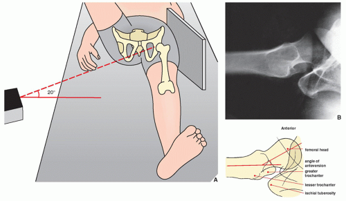
FIGURE 8.7 Groin-lateral view. (A) For the groin-lateral view of the hip, the patient is supine with the affected extremity extended and the opposite leg elevated and abducted. The cassette is placed against the affected hip on the lateral aspect, and the central beam is directed horizontally toward the groin with approximately 20 degrees cephalad angulation. (B) The radiograph obtained in this projection provides almost a true lateral image of the femoral head, thereby allowing evaluation of its anterior and posterior aspects. It also demonstrates the anteversion of the femoral neck, which normally ranges from 25 to 30 degrees.


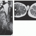
FIGURE 8.8 CT of the sacroiliac and hip joints. (A) CT section at the level of S2 demonstrates the true (synovial) sacroiliac joints. (B) In this section through the hip joints, the relation of the femoral heads to the acetabula can be evaluated sufficiently. The pubic bone and the pubic symphysis are also well delineated.


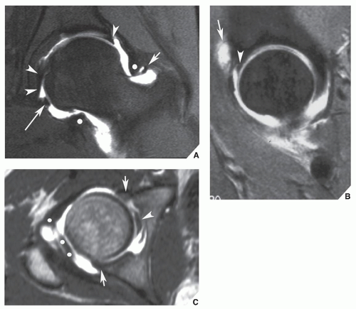
FIGURE 8.9 Normal MRa of the hip. (A) Coronal T1-weighted fat-saturated MRa demonstrates the normal superior labrum (single arrowhead), the ligamentum teres (double arrowheads), the orbicular zone (white dots), the transverse ligament (long arrow), and the superior retinaculum (short arrow). (B) Sagittal T1-weighted fat-saturated MRa demonstrates the superior labrum (arrowhead) and a small amount of contrast material in the iliopsoas bursa (arrow), a feature seen in about 15% of the normal population. (C) Axial T1-weighted MRa demonstrates the ligamentum teres (arrowhead), the orbicular zone (white dots), and the anterior and posterior labrum (arrows).


TABLE 8.1 Standard and Special Radiographic Projections for Evaluating Injury to the Pelvis, Acetabulum, Sacrum, and Proximal Femur


Projection


Demonstration


Anteroposterior


Angle of femoral neck


Radiographic landmarks (lines) relating to acetabulum:


Iliopubic (iliopectineal)


Ilioischial


Teardrop


Acetabular roof


Anterior acetabular rim


Posterior acetabular rim


Varus and valgus deformities


Avulsion fractures


Malgaigne fracture


Fractures of


Ilium (Duverney)


Ischium


Pubis


Sacrum (in some cases)


Femoral head and neck


Dislocations in hip joint


With 30-35 degrees cephalad angulation (Ferguson) (or posteroanterior with or without 25-30 degrees caudal angulation)


Fractures of


Sacrum


Pubis ramus


Ischium


Injury to sacroiliac joints


Oblique (Judet views)


Anterior (internal)


Iliopubic line


Fractures of


Anterior (iliopubic) column


Posterior acetabular rim


Posterior (external)


Quadrilateral plate


Fractures of


Posterior (ilioischial) column


Anterior acetabular rim


Frog-Lateral


Fractures of


Femoral head and neck


Greater and lesser trochanters


Groin-Lateral


Angle of anteversion of femoral head


Anterior and posterior cortices of femoral neck


Ischial tuberosity


Rotation and displacement of femoral head in subcapital fractures


TABLE 8.2 Ancillary Imaging Techniques for Evaluating Injury to the Pelvis, Acetabulum, and Proximal Femur


Technique


Demonstration


CT (including 3D CT)


Position of fragments and extension of fracture line in complex fractures, particularly of pelvis, acetabulum, and sacrum


Weight-bearing parts of joints


Sacroiliac joints


Intraarticular fragments


Soft-tissue injuries


Concomitant injury to ureters, urinary bladder, and urethra


MRI


Soft-tissue injuries, including various tendon abnormalities, compressive and entrapment neuropathies (piriformis syndrome), and Morel-Lavallée lesion


Posttraumatic osteonecrosis


Occult fractures


Bone contusions (trabecular microfractures)


CT-angiography


Injury to the vascular system


Radionuclide imaging (scintigraphy, bone scan)


Occult fractures


Stress fractures


Posttraumatic osteonecrosis


IVP


Cystourethrography


Concomitant injury to ureters, urinary bladder, and urethra


Angiography (arteriography, venography)


Injury to vascular system


CT, computed tomography; 3D, three-dimensional; IVP, intravenous urography; MRI, magnetic resonance imaging.


FIGURE 8.10 Spectrum of radiologic imaging techniques for evaluating injury to the pelvic girdle. The radiographic projections or radiologic techniques indicated throughout the diagram are only those that are the most effective in demonstrating the respective traumatic conditions. #Replaced almost completely by CT. CT, computed tomography; AP, anteroposterior.


FIGURE 8.11 Stable pelvic fractures. (Modified from Dunn AW, Morris HD. Fractures and dislocations of the pelvis. J Bone Joint Surg [Am] 1968;50A:1639-1648.)


Injury to the Pelvis and Acetabulum

The pelvis is a nearly rigid ring essentially comprising three elements: the sacrum and two paired lateral components, each composed of the ilium, the ischium, and the pubis. Because of this configuration and the interrelationship of its components, identification of an apparently solitary fracture should not end the process of radiographic examination. The pelvis should be scrutinized carefully for other fractures of the ring or diastasis in the sacroiliac joints or the pubic symphysis (see Fig. 4.7).


Classification of Pelvic Fractures

Various classification systems have been proposed not only to identify the distinctive appearances of pelvic injuries as an aid to radiographic recognition and diagnosis but also to categorize such injuries as an aid to orthopedic management and prognosis. The latter point is particularly important in pelvic fractures because of the inherent instability of the structures composing the pelvic girdle, their integrity depending entirely on ligamentous support and the stabilizing influence of the sacroiliac joints. Thus, pelvic fractures can be grouped according to whether they significantly detract from the stability of the pelvic ring, with the orthopedic management and prognosis of those fractures identified as stable (Fig. 8.11) differing considerably from that of unstable fractures (Fig. 8.12).


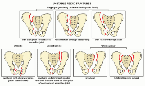
FIGURE 8.12 Unstable pelvic fractures. (Modified from Dunn AW, Morris HD. Fractures and dislocations of the pelvis. J Bone Joint Surg [Am] 1968;50A:1639-1648.)

Systems that classify pelvic injuries for the purpose of radiographic diagnosis and orthopedic management using categories other than stable and unstable have also been suggested. Pennal, Tile, and colleagues have elaborated a system based on the direction of the force that produces
pelvic injuries. They identified four patterns of force as underlying mechanisms of injury that produce distinctive radiographic appearances:


  • Anteroposterior compression, in which the force vector in the anteroposterior or posteroanterior direction produces vertically oriented fractures of the pubic rami and disruption of the pubic symphysis and sacroiliac joints, which often results in bilateral pelvic “dislocation” (sprung pelvis, “open book” injury).


  • Lateral compression, in which the lateral force vector often results in horizontally or coronally oriented fractures of the pubic rami, compression fractures of the sacrum, fractures of the iliac wings, and central dislocation in the hip joint as well as varying degrees of pelvic instability caused by displacement or rotation of one or both hemipelves, depending on whether the compressive force is applied more anteriorly or more posteriorly.


  • Vertical shear, in which the inferosuperiorly oriented disruptive force, delivered to one or both sides of the pelvis lateral to the midline often as a result of a fall from a height, frequently produces vertically oriented fractures of the pubic rami, sacrum, and iliac wings. Because of significant ligamentous disruption, this type of force is associated with injuries producing severe pelvic instability.


  • Complex patterns, in which at least two different force vectors have been delivered to the pelvis, the patterns produced by anteroposterior and lateral compression being the most commonly encountered.

This system, which corresponds to the more traditional categorization of pelvic fractures into stable and unstable, has practical value in allowing sufficient evaluation of pelvic injuries to be made on the anteroposterior projection in patients requiring immediate surgical intervention when CT scans would be impractical to obtain. It also provides correlations between the type of force delivered to the pelvis and the concomitant ligamentous and pelvic-organ injury that can be expected. In anteroposterior compression-type injuries, for example, the anterior sacroiliac ligaments, the sacrotuberous-sacroiliac ligament complex, and the symphysis ligaments are damaged. This type of injury may also be associated with urethral and urinary bladder rupture and damage to the pelvic blood vessels. In lateral compression injuries, rupture of the posterior sacroiliac ligament and/or the sacrospinous-sacrotuberous ligament complex may result. Injury to the urinary tract may or may not be present. In vertical shear injuries, the ipsilateral posterior and anterior sacroiliac, the sacrospinous-sacrotuberous, and the anterior symphysis ligaments are usually ruptured. Vertical shear injuries are frequently accompanied by damage to the sciatic nerve and pelvic blood vessels, often resulting in massive hemorrhage. The discussion that follows, however, focuses on the more traditional pedagogic categories of pelvic trauma.


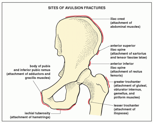
FIGURE 8.13 Sites of avulsion fractures.


Fractures of the Pelvis


Avulsion Fractures

Usually involving the anterosuperior or anteroinferior iliac spine or the ischial tuberosity, avulsion fractures, which are classified as stable fractures (Fig. 8.13; see also Fig. 8.11), most commonly occur in athletes as a result of forcible muscular contraction: the sartorius muscle and tensor fasciae latae in avulsion of the anterosuperior iliac spine, the rectus femoris muscle in avulsion of the anteroinferior iliac spine, the hip rotators in avulsion of greater trochanter, the iliopsoas in avulsion of the lesser trochanter, the adductors and gracilis in avulsion of pubic bone, and the hamstrings in avulsion of the ischial tuberosity. Most fractures of these structures are apparent on a single anteroposterior radiograph of the pelvis (Fig. 8.14). However, confusion in diagnosis may arise when healing occurs by exuberant callus formation, at which time or after full ossification such fractures may be mistaken for neoplasms. Another entity that may mimic avulsion injury to the pelvis is the so-called pelvic digit, a congenital anomaly characterized by a bone formation in the soft tissue about the pelvic bones (Fig. 8.15).


FIGURE 8.14 Avulsion fractures. A 16-year-old boy was injured during an athletic activity. (A) Anteroposterior radiograph of the pelvis shows a crescent-shaped fragment adjacent to the lateral aspect of the iliac wing (arrow), which represents the avulsed apophysis of the anterosuperior iliac spine. (B) Anteroposterior radiograph of the hip in a 26-year-old runner clearly demonstrates avulsion of the ischial tuberosity. (C) As a sequela of avulsion of the ischial tuberosity and injury to the soft tissue in the region, a 28-year-old athlete had ossification of the obturator externus muscle.


Malgaigne Fracture

This unstable injury, involving one hemipelvis, most commonly consists of unilateral fractures of the superior and inferior pubic rami and disruption of the ipsilateral sacroiliac joint (see Fig. 8.12). In the variants of this type of injury, the unilateral fractures of the pubic rami may be accompanied by a fracture through the sacral wing near the sacroiliac joint or through the ilium (see Fig. 8.12). Separation of the pubic symphysis may coexist with such injuries, and cephalad or posterior displacement of the entire hemipelvis may occur. The Malgaigne fracture, which is recognized clinically by shortening of the lower extremity, is readily demonstrated on the anteroposterior radiograph of the pelvis (Fig. 8.16).


Miscellaneous Pelvic Fractures

Injuries other than the Malgaigne fracture are also easily evaluated on radiographs of the pelvis in the standard and special projections or on CT examination. The Duverney fracture is a stable fracture of the wing of the ilium without interruption of the pelvic ring (see Fig. 8.11). The straddle fracture (see Fig. 8.12) consists of comminuted fractures of both obturator rings (i.e., all four ischiopubic rami). In one third of patients with this unstable fracture, bladder rupture or urethral injuries occur. The bucket-handle or contralateral double vertical fracture involves the superior and inferior ischiopubic rami on one side combined with fracture about or disruption of the sacroiliac joint on the opposite side (see Fig. 8.12). Fractures of the sacrum (see the following text), which may be either transversely or vertically oriented (see Figs. 8.11 and 8.27, 8.28, 8.29, 8.30), may occur alone or, more often, in association with other pelvic injuries, such as the so-called pelvic dislocations. The latter are characterized by disruption in one or both sacroiliac joints (unilateral or bilateral dislocation) associated with separation of the pubic symphysis (Fig. 8.17; see also Fig. 8.12). The anteroposterior projection obtained with 30 degrees cephalad angulation or CT is helpful in disclosing sacral fractures, which are frequently overlooked.


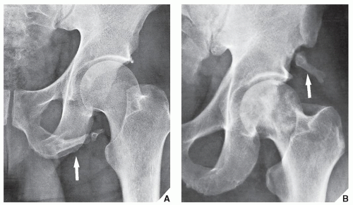
FIGURE 8.15 Pelvic digit. A rare congenital anomaly, the pelvic digit may occasionally be mistaken for avulsion fracture. (A) Anteroposterior radiograph of the left hip shows a finger-like, jointed structure attached to the caudal portion of the left ischium (arrow). (B) Anteroposterior radiograph of the hip in a 55-year-old man with no history of trauma demonstrates a well-formed digit at the site of the anteroinferior iliac spine (arrow). (From Greenspan A, Norman A. The “pelvic digit”—an unusual developmental anomaly. Skeletal Radiol 1982;9:118-122.)


Fractures of the Acetabulum

Evaluation of the acetabulum on conventional radiographs may be difficult because of obscuring overlying structures. If acetabular fracture is suspected, then radiographs in at least four projections should be obtained: the anteroposterior view of the pelvis, the anteroposterior view of the hip, and the anterior and posterior oblique (Judet) views. Radiography may also need to be supplemented by CT, as discussed previously.

As an aid in recognizing the presence of abnormality on the anteroposterior projection of the pelvis and hip, Judet, Judet, and Letournel have identified six lines relating to the acetabulum and its immediately

surrounding structures (Fig. 8.18). Fracture of the acetabulum usually distorts these radiographic landmarks, allowing a diagnosis to be made on the anteroposterior projection, but an accurate and complete evaluation of the fracture requires that oblique views be obtained (Fig. 8.19). As mentioned, the anterior (internal) oblique projection demonstrates the iliopubic column and the posterior lip of the acetabulum (see Fig. 8.4), and the posterior (external) oblique view images the ilioischial column and the anterior rim of the acetabulum (see Fig. 8.5). These projections, together with the division of the pelvic bone into anterior and posterior columns (Fig. 8.20), provide the basis for the traditional classification of acetabular fractures. This classification has been modified by Letournel to include the following types of fractures (Fig. 8.21) :

Only gold members can continue reading. Log In or Register to continue

Stay updated, free articles. Join our Telegram channel

Jul 24, 2016 | Posted by in MUSCULOSKELETAL IMAGING | Comments Off on Lower Limb I: Pelvic Girdle, Sacrum, and Proximal Femur

Full access? Get Clinical Tree

Get Clinical Tree app for offline access