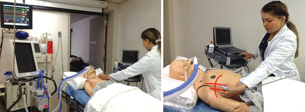
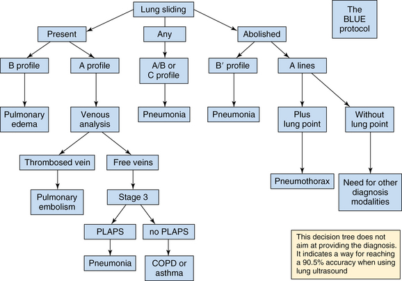
24 Use of a microconvex transducer with a 4- to 8-MHz frequency range is recommended. In general, the microconvex transducer is used for the evaluation of the lung and the pleural space, but a high-frequency probe also can be used for the specific diagnosis of a pneumothorax when using ultrasound for vascular access. The intubated patient is almost always examined in the semirecumbent or supine position (Figure 24-1). Figure 24-1 Patient positioning. The intubated patient is almost always examined in supine position. Check all six anatomic regions: upper and lower sections of the anterior, lateral, and posterior chest wall. 1. Expose the thorax and place ultrasound gel on the six regions of the chest. 2. Start the examination with the transducer in the first intercostal space in the upper anterior region of the chest, with the ultrasound marker pointing toward the head of the patient. 3. Continue the examination by sliding the ultrasound probe in a longitudinal direction, from the head toward the toes of the patient. 4. Visualization is performed in each intercostal space, concentrating on the image between the two rib shadows, and should last for at least one complete respiratory cycle. 5. Repeat the process in the lateral and posterior chest walls in the same manner. 6. The following lung findings are evaluated: 7. In addition to lung findings, the following can be evaluated to supplement the examination: In 2008, Daniel Lichtenstein evaluated the potential of lung ultrasound in diagnosing acute dyspnea.1 Ultrasonography was performed on patients with acute respiratory failure, comparing lung ultrasonography results on initial presentation with the final diagnosis by the ICU team. The study developed the BLUE Protocol (Bedside Lung Ultrasound in Emergency Protocol) as a diagnostic tool (Figure 24-2). The following patterns and profiles were established: Figure 24-2 The BLUE Protocol: A decision tree using lung ultrasonography to guide diagnosis of severe dyspnea. (From Lichtenstein D, Mezière G: Relevance of lung ultrasound in the diagnosis of acute respiratory failure: the BLUE Protocol. Chest 134(1):117-125, 2008.) 1. Predominance of A-lines in the presence of lung sliding indicated an exacerbation of asthma or chronic obstructive pulmonary disease (COPD) (89% sensitivity and 97% specificity). 2. Multiple, anterior diffuse B-lines in the presence of lung sliding indicated pulmonary edema (97% sensitivity and 95% specificity). 3. Normal anterior profile in the presence of a deep venous thrombosis indicated pulmonary embolism (81% sensitivity and 99% specificity).
Lung ultrasound: Protocols in acute dyspnea
Instrumentation and technique
Related posts:
 Ultrasound-guided peripheral intravenous access
 Integrating picture archiving and communication systems and computerized provider order entry into the intensive care unit: The challenge of delivering health information technology–enabled innovation
 Endobronchial ultrasound: (CONSULTANT-LEVEL EXAMINATION)
 Ultrasonography in circulatory failure
 Evaluation of right ventricular function in the intensive care unit by echocardiography: (CONSULTANT-LEVEL EXAMINATION)
 Procedural ultrasound for surgeons: (CONSULTANT-LEVEL EXAMINATION)
![]()
Stay updated, free articles. Join our Telegram channel

Full access? Get Clinical Tree


Lung ultrasound: Protocols in acute dyspnea
