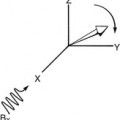
Magnetic Resonance Images
Objectives
At the completion of this chapter, the student should be able to do the following:
Key Terms
What is an Image?
In the most general sense an image is a mental picture. It may be a visual image based on direct observation, such as the viewing of the Grand Canyon, or an imaginary image, such as the duck elicited by the sound of the oboe in Prokofiev’s Peter and the Wolf. An image can also be abstract, such as the famous painting by Tanner (Figure 15-1).
Medical images generally fall into the category of visual images; they attempt to represent real objects accurately. A brief discussion of some basic concepts of visual images is helpful in explaining magnetic resonance (MR) images.
Visual Images
All visual images are initially detected by the eye and are the result of stimulation of receptor cells in the retina by electromagnetic radiation in the visible region of the spectrum. The two types of receptor cells are rods and cones. These receptor cells can be considered digital detectors that are stimulated by the input of light photons. They respond to such stimulation with an output of discrete electrical impulses called action potentials.
Each retinal receptor is arranged by the optical structure of the eye to be sensitive to light coming from a specific region of the visual field and to characterize the nature of that light as to intensity and color. This initial information about the source and character of light is then relayed through complex visual pathways to the occipital lobe, where the most complex processing and analysis of these data are performed (Figure 15-2).
In the occipital lobe, the myriad of discrete data concerning light impinging on the retina is reconstructed into the perceived image. Although the final image is often considered to be a continuum of information about light, it is initially detected, processed, and conceived as a high-resolution digital image.
Optical Receptors
When light arrives at the retina, it is detected by the rods and the cones. Rods and cones are small structures; there are more than 100,000 of them per square millimeter of the retina.
The cones are concentrated on the center of the retina in an area called the fovea centralis. On the other hand, rods are most numerous on the periphery of the retina. There are no rods at the fovea centralis.
The rods are very sensitive to light and are used in dim-lighting situations. The threshold for rod vision is approximately 10−6 ml. However, cones are less sensitive to light; their threshold is only 5 × 10⁻³ ml, but they can respond to intense light levels, whereas rods cannot. Consequently, cones are used primarily for daylight vision, called photopic vision, and rods are used for night vision, called scotopic vision.
This aspect of visual physiology explains why dimly lit objects are more readily viewed if they are not looked at directly. Astronomers and radiologists are familiar with the fact that a dim object can be seen better if viewed peripherally, in which case rod vision dominates.
The ability of the rods to visualize small objects is much worse than that of the cones. This ability to perceive fine detail is called visual acuity. Cones are also much more able than rods to detect differences in brightness levels. This property of vision is termed contrast perception. Furthermore, cones are sensitive to a wide range of wavelengths of light.
 Cones perceive color; rods are essentially color-blind.
The cones of the eye are color receptors of three types, each stimulated by relatively narrow bandwidths of light generally referred to as red, blue, and green. They are sensitive to brightness (number of light photons), hue (wavelength), and saturation (ratio of monochromatic to white light). The human eye can detect approximately 2000 different color combinations or hues compared with perhaps only 20 shades of gray.
Therefore color images contain significantly more information than black and white images. Imaging techniques such as magnetic resonance imaging (MRI) intrinsically contain more information and are more naturally adaptable to color imaging.
In a standard clinical setting, when someone views a 14 × 17 inch chest radiograph at 50 cm, the eye can spatially resolve 0.5 to 1 mm, 20 shades of gray, and 1000 different colors. Optimal medical imaging should take maximum advantage of this basic physiologic capability. Functional MRI (fMRI) is a first step in this direction.
Pattern Recognition
The spatial map of light that is a visual image does not have any intrinsic intellectual significance. The intellectual value of such an image primarily depends on the mental comparison of an image with the large library of images stored within the brain and the subsequent evaluation of the meaning of the image.
Most medical imaging is a process of pattern recognition, which basically involves comparing one image pattern with another. Evaluation for similarities and differences with known patterns results in a “best fit” conclusion or “most likely” diagnosis (Figure 15-3).
This leads to the inescapable fact that a major factor in diagnostic efficiency is the observer’s development of a large image memory bank, which at least partially comes from viewing many images. Such a memory bank is currently implemented and supplemented in CAD (computer-assisted detection).
Regardless of the significance of an image, visual images can be evaluated on the basis of spatial resolution and contrast resolution. That is, the perception and interpretation of an image depend on the location of light photons and the differences in character of those photons.
Location and Character
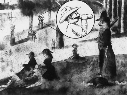
An image consists of discrete spatial points, each having different light characteristics. This concept was appreciated and emphasized by a group of Postimpressionist painters called pointillists, who painted pictures made up of dots of different colors. When they are viewed at close range, the dots are obvious but what they represent is not, but at a distance, the dots appear to merge into a continuum of space and color forming shapes and patterns.
The painting shown in Figure 15-4 is patterned after the work of a famous pointillist, Georges Seurat, and illustrates this concept. When it is viewed at a distance, the painting is of a picnic in the Texas hill country and elicits the psychological impression of such a real scene. When it is viewed at close range, the painting loses its totality and becomes simply a matrix of dots.
Such artists called these dots points, but these dots are perfectly analogous to what we call pixels. Each pixel has a unique location and character. The location is the position of each pixel in relationship to others. The character is represented by brightness, color, or both.
Most medical images differ from paintings in what the character of the pixel means. When an object is directly viewed, the pixel character is the actual brightness and wavelength of light reflected to the eye. When a representational image is viewed, the pixel character still primarily reflects the light incident onto the eye, but this initial character of light represents or stands for something else. In a realistic painting, the pixel character represents the visual light characteristics of actual objects.
 Medical images are representational.
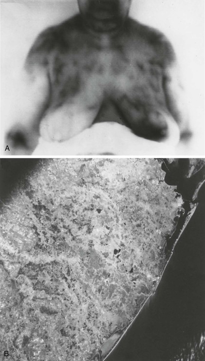
Medical images represent something else and are not realistic. They do not attempt to copy a real, directly visualized object. For example, in the infrared images shown in Figure 15-5, the pixel character represents the radiant heat of an object, a feature that cannot be seen directly.
Equally representational is the temperature map shown in Figure 15-6 in which the various pixels stand for the temperature of a region rather than any geographic feature; such a representational image can be made whenever any parameter can be measured as a function of location.

Representational Images
Medical images are representational because the character of the pixel does not attempt to reflect the actual visual light feature of the tissue, but rather, some other physical parameter that has been measured and transformed into the image. The character of a pixel can be made to stand for essentially any measurable quantity that can be spatially defined. Examples include electron density maps (radiographs), radioactive decay maps (radionuclide images), and hydrogen concentration maps (MR images).
Another obvious difference between most directly observed images and medical images is that medical images are of the interior of a body rather than the surface. The primary clinician does the medical imaging of the surface of the body by direct observation, whereas the radiologist images the inside of the body.
Radiographic Images
Imaging the inside of the body was a major problem in early medicine. Before Roentgen’s discovery of x-rays in 1895, the only way medical images of the interior of the body were made was by direct visualization. This obviously required cutting into the subject and viewing the organ or tissue of interest in the form of traditional medical illustration (Figure 15-7).

With the discovery of x-rays, it became possible to make images of the interior of the body in a relatively noninvasive fashion. Radiographic images have features comparable to the representational images discussed. A radiographic image has pixels (groups of silver grains) with unique location and concentration differences that account for pixel character or brightness.
The basic difference between the radiographic image and a direct visual image of the body is in the devices that measure pixel character or the detectors. In the case of direct visualization, the detectors are the rods and cones of the retina. In the case of a radiograph, the detectors are the silver halide grains of the film emulsion that are sensitive to short-wavelength electromagnetic radiation.
The number of x-rays detected determines pixel character. The amount of x-rays absorbed by the body is physically determined by the x-ray attenuation coefficient, which is principally related to the density of electrons in different tissues.
 A radiograph is essentially an electron density map of the body.
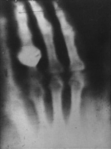
However, a radiograph does not relay any numerical information about the electron density (Figure 15-8). The informed observer knows only that the blackness or whiteness on the image is related to electron density. The whiter the image, the higher is the electron density.
This gray scale information is very useful, even if the physics of x-ray interaction and electron density are not considered. This concept is used empirically to evaluate the internal structure of the anatomy.
Radiographic contrast resolution among tissues is determined principally by differences in electron density. For example, bone has a much higher electron density than soft tissue and is therefore highly contrasted to soft tissues on a radiograph. The evaluation of bone remains one of the main uses of radiography.
Radiographs differentiate among tissues and therefore report gross anatomy. However, radiographs can also relate pathologic anatomy by showing abnormal gross anatomy, as reflected by deformities, distortions, absence, enlargement, and reduction of organs and tissues.
In addition to providing information about normal and abnormal gross anatomy, radiographs can provide some information about physiology, such as the healing of fractures reflected by calcium deposition in callus formation or the abnormal deposition of calcium in the basal ganglia in patients with hypercalcemia.
Magnetic Resonance Images
Unfortunately, most anatomical or physiologic implications of radiographic images are indirect because there is no intrinsic biological significance to electron density. The clinical significance of an x-ray image is based on the recognition of patterns from accumulated experience of empirically evaluated radiographs.
Essentially, all forms of medical imaging use the substitution of some primary physical detector system other than the human visual system. The visual system becomes a secondary receptor that interprets the representational images created by the primary receptor system.
All medical image detector systems create a spatially defined map of the human body as a function of some particular physical parameter. Such medical images, then, are really three-dimensional images. Two dimensions define pixel location, and the third dimension, represented by brightness or color, contains information about the physical character of the tissue.
 An MR image is essentially a map of the magnetic characteristics of nuclei within the atoms in the body.
The physical character of the pixels in an MR image is multidimensional. The numerical value of an MRI pixel is principally determined by the MRI parameters: proton density (PD), spin-lattice relaxation time (T1), and spin-spin relaxation time (T2).
Secondary determinants of MRI pixel values are chemical shift, magnetic susceptibility, and motion. The weighting given to each of these parameters can vary greatly depending on the timing of the radiofrequency (RF) pulses and the gradient magnetic fields.
Image Evaluation Criteria
Spatial Criteria
The primary value of pixel location is to display geometry or gross anatomy. The display of gross anatomy requires not only appropriate pixel positioning but also adequate differences in pixel character to produce sufficient contrast between tissues to allow their separation and recognition.
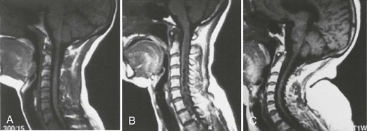
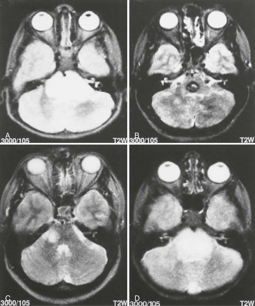
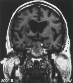
However, most evaluation of gross anatomy is geographic and therefore relatively independent of the imaging technique. For instance, once the lateral ventricles have been displayed on an image (Figure 15-9), the character of the pixels becomes largely irrelevant.
 The three principal spatial criteria are size, shape, and position.
The relevant factors for the evaluation of the gross anatomy of the lateral ventricles or any other structure are basically the geometric factors: size, shape, and position. In the viewing of the image, the size, shape, and position of the lateral ventricles are referenced to normal, which is based on experience.
Evaluation of the spatial aspects of an image is relatively straightforward. The specific possibilities in terms of size are normal, large, and small (Figure 15-10).