Partially Parallel Magnetic Resonance Imaging
Objectives
At the completion of this chapter, the student should be able to do the following:
Key Terms
In this chapter, the basic approaches to parallel imaging—and there are several—will be introduced. Parallel imaging is a relatively new type of MR imaging that exploits features of phased array RF coils to significantly increase the speed of MR image acquisition.
Recall that phase array coils are groups of receiver coils that each sense the nuclear magnetization from a small portion of the patient. The images from the individual coils are stitched together to form an overall image of a large body part that has both good SNR and relatively good image uniformity.
Parallel imaging is a method for encoding the data which is generally compatible with many types of pulse sequences. It is particularly useful in high-field MRI systems (3 tesla and above) because it allows the use of fewer RF pulses than conventional imaging methods, thus reducing the potential for tissue heating. We shall explore the hardware requirements and some of the new imaging parameters that are important for parallel imaging; particularly the relationship between the geometry factor and SNR in parallel imaging.
We shall also take a look at various clinical applications of parallel imaging. A comprehensive account of what parallel imaging can do is not necessary for operating an MR imaging system, but in the description below is aimed a giving the reader a flavor of the advantages of parallel imaging and some situations in which it might be used.
General Description of Parallel Imaging
The key elements of partially parallel imaging (PPI) are, first of all, it uses information obtained from arrays of RF coils sampling data in parallel. Since each coil element is located at a different position, this information can be used to perform some portion of spatial encoding that was usually done by the gradient fields, particularly the phase-encoding gradient field.
Imaging time is reduced because the phase-encoding process is the slowest part of image formation in MRI. So fundamentally PPI speeds up the MR image acquisition process and it does this without needing faster switching gradients, avoiding the tissue stimulation issues they can be caused by using additional RF pulses. In fact, parallel imaging can actually reduce the number of RF pulses so that the RF power deposited is not a big concern.
Now there are two fundamental approaches to parallel imaging. The first is an image based method in which the image is basically reconstructed from the signals detected by each coil element in the phased array with a reduced field of view and then merged. The most common implementation of this is known as SENSE, which stands for sensitivity encoding.
The second approach to producing parallel images is the k-space based approach. In this method the raw image data are analyzed and an interpolation algorithm is used to interpolate missing data in k-space before the Fourier transformation occurs. The algorithm known as GRAPPA is most commonly implemented on clinical MRI systems now, although other variations of this method are under development.
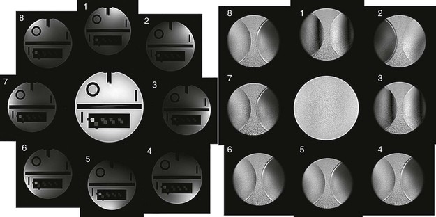
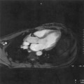
You may ask, “What’s the difference between parallel imaging and just regular imaging with phased array coils?” Well, in Figure 22-1 two examples of the images obtained from an eight-channel phased array coil are shown. On the left, there is a typical phased array type configuration in which each phased array coil picks up different signals from different parts of the phantom and then these images are added together to form an image of the phantom that looks much more uniform than you can get from any individual surface coil.
With parallel imaging, to speed up the imaging process, data are acquired for a smaller field of view. For instance, if you want to double the imaging speed, then use an FOV in phase-encoding direction that is half of what you really want. Obviously, this takes half of the imaging time but it also creates problems.
A reduced data set has been acquired with each coil and so, as we shall find out in Chapter 27, the image will have a wraparound or aliasing artifact that appears in the phase-encoding direction in each individual image where the top part of the patient is wrapped into the bottom part of the image. Now, the whole trick of the parallel imaging process is to unwrap these aliased images and put them back together into one image. We get increased imaging speed from this method, but we will also have lower SNR since we are acquiring real data for fewer lines in k-space.
 Partially parallel imaging methods increase imaging speed by reducing the number of phase-encoding lines in k-space that need to be acquired.
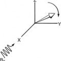
A useful analogy is to compare parallel MRI to x-ray CT where projections of data are used for both cases. But in parallel MRI, the projections come from each of the RF coils and their spatial position and their sensitivities are a function of spatial position (Figure 22-2), so this is a very simple analogy that has some limitations. The first major problem is that in general there is no fixed geometry between the coils and the patient. Second, there is coupling between the patient—we can think of the patient as being composed of conductive bags of salt water—and the coil array. Third, the setup changes from study to study and may even change sometimes within a given study.
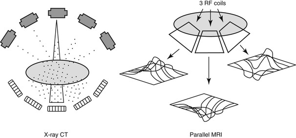
There is also the fact that the surface coils themselves receive MR signals in an inherently nonuniform fashion. These factors make it seem like it’s going to be very difficult to determine spatial properties of the signals based on the geometry and locations of the coils.
The key to parallel imaging is to acquire a little bit of extra data to get the information needed regarding the spatial distribution of RF coils’ sensitivity. One way to do this is by electromagnetic modeling of the coils’ properties applied to phantom measurements. However, because every patient differs and the way every patient couples with a coil differs, this approach is practical only for designing the RF coils used for parallel imaging. In the general clinical situation, extra data collection on the patient, either before or during the imaging, is used to gain a priori knowledge of how the patient and coil work together before parallel imaging reconstruction is performed.
 Parallel imaging requires that extra data on the RF coils’ signal sensitivity as a function of position in the patient be obtained before image reconstruction.
Image Based Parallel Imaging Reconstruction
SENSE is the primary image based form of PPI which is, in some form or another, offered on all the high-end imaging systems of all the major manufacturers. In the common implementation of SENSE, sensitivity profiles are determined for each element of the phased array coil. Measuring the coil sensitivity profiles gives the system the additional information necessary to constrain the reconstruction process so that it can be accurate and efficient.
The two basic methods used for parallel imaging differ in their approaches to obtaining the data to solve the problem of recovering the spatial information from the set of RF coils. The coil sensitivity profiles contain the data that describes the spatial dependencies of each RF coil.
Related posts:


Stay updated, free articles. Join our Telegram channel

Full access? Get Clinical Tree


