Perfusion Imaging
Objectives
At the completion of this chapter, the student should be able to do the following:
Key Terms
The previous chapter described the imaging of large vessels with magnetic resonance (MR) techniques, magnetic resonance angiography (MRA). This chapter describes techniques for functional magnetic resonance imaging, imaging blood microcirculation, perfusion. The following chapter describes magnetic resonance imaging (MRI) at an even lower anatomical level, diffusion.
 Both perfusion imaging and diffusion imaging are the bases for functional magnetic resonance imaging (fMRI).
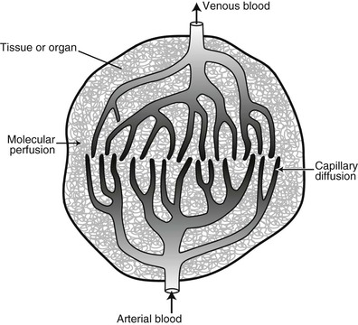
Blood flows through the vascular tree into smaller and smaller vessels. Capillaries are the smallest vessels, and they are responsible for delivering oxygen and nutrients to tissues. Imaging blood flow in capillaries is perfusion imaging.
Blood delivered to tissue, arterial blood, is oxygenated. Venous blood is removed from tissue through a similar capillary network as deoxygenated blood (Figure 24-1).
 Perfusion is the flow of blood through the capillary network.
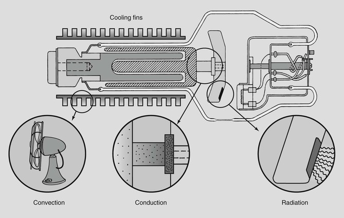
The x-ray tube and a hot water radiator can serve to distinguish perfusion from diffusion. In an x-ray tube, the anode heat is dissipated by conduction to the rotor, convection from the housing, and radiation from the anode (Figure 24-2). In a room-warming radiator, hot water is conducted through the coils of the radiator where heat is transferred to air molecules, which are convected through the room. In each of these examples, conduction is analogous to perfusion; convection is analogous to diffusion.
There are two general approaches to perfusion imaging: the use of exogenous contrast agents and the use of endogenous contrast agents (Box 24-1).
Exogenous Magnetic Resonance Imaging
The use of exogenous contrast agents for studying brain perfusion has been practiced for some time with considerable success. Single photon emission computed tomography (SPECT) with 133Xe, positron emission tomography (PET) with 15O, and x-ray computed tomography (CT) with an iodinated contrast agent provide the basic approach to what has become exceptionally effective MR perfusion imaging.
Related posts:


Stay updated, free articles. Join our Telegram channel

Full access? Get Clinical Tree


