Chemical Shift and Magnetization Transfer
Objectives
At the completion of this chapter, the student should be able to do the following:
Key Terms
The gyromagnetic ratio for hydrogen is 42 MHz/T. Therefore, in a 1 T B0 field, hydrogen nuclei precess with a resonant frequency of 42 MHz. Table 17-1 summarizes hydrogen proton resonant frequency at several other B0 values.

For a magnetic resonance imaging (MRI) signal to be obtained from tissue, the transmitted radiofrequency (RFt) must be at the prescribed resonant frequency, say 42 MHz for a 1 T MRI system, to excite the proton spins. However, this situation applies only to proton spins in mobile water molecules, so-called free water.
Although water makes up approximately 80% of all tissue molecules, hydrogen is only approximately 60% of all tissue atoms. Nevertheless, the hydrogen in water predominates and is the principal source of the magnetic resonance (MR) signal.
 Sixty percent of the atoms of human tissue are hydrogen atoms.
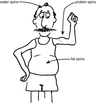
However, proton spins in other tissue atoms also contribute to the MR signal if they are excited at the proper resonant frequency. Many schemes have been proposed for compartmentalizing body tissue for the purpose of MRI. The three-compartment model (Figure 17-1) is the simplest and all that is necessary for the discussion of magnetization transfer (MT)—water protons, fat protons, and macromolecular protons. Before proceeding to MT, consider again chemical shift because they are related.
Chemical Shift
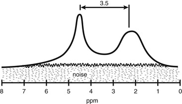
A revisit to the discussion of the parts per million (ppm) scale in Chapter 7 will show that ppm is a relative value compared with the standard molecule tetramethylsilane (TMS). TMS is assigned an arbitrary value of 0 on the ppm scale. Water protons are shifted downfield 4.7 ppm and those of fat, about 1.2 ppm. This is shown in the spectrum of Figure 17-2.
The reason that the chemical shift is expressed in ppm is that the shift will increase as B0 increases. Thus, in a 1-T B0 field, water protons will resonate at 300 Hz lower than TMS and fat protons have a resonant frequency about 51 Hz below that of TMS. However in a 3 T B0 field, water protons will resonate at 900 Hz lower than TMS and fat protons will resonate about 153 Hz below TMS. Using the ppm scale allows the chemical shift to be more easily related between MRI systems with different B0 fields.
All hydrogen in water is bound to oxygen in the same manner. Therefore the water peak is narrow and intense. Hydrogen in fat is bound in many different ways, and this causes the fat peak to be broader and less intense. It is actually the result of many individual spectral lines, each representing a different type of chemical bond.
The resonant frequency for fat differs from that for water because of very small differences in the local magnetic field. Both molecules may reside in an equal B0 field, but the local magnetic field is slightly different and determined by the configuration of electrons in the bound molecule. This phenomonon is known as “electron shielding.” Hydrogen spins and other nuclear spins in the same molecule can also cause smaller disturbances of the local magnetic fields, which is manifested in the J-coupling mentioned in Chapter 16.
 Free water protons and fat protons have a resonance frequency separation of about 3.5 ppm.
Because fat and water proton spins are at 4.7 and 1.2 ppm, respectively, their resonant frequency separation is (4.7 − 1.2 = 3.5) 3.5 ppm. At 0.5 T, the frequency difference is small. However, at 1.5 T and higher, the frequency difference can result in a significant artifact.