Mandible and Muscles of Mastication Axial 1

Normal Anatomy
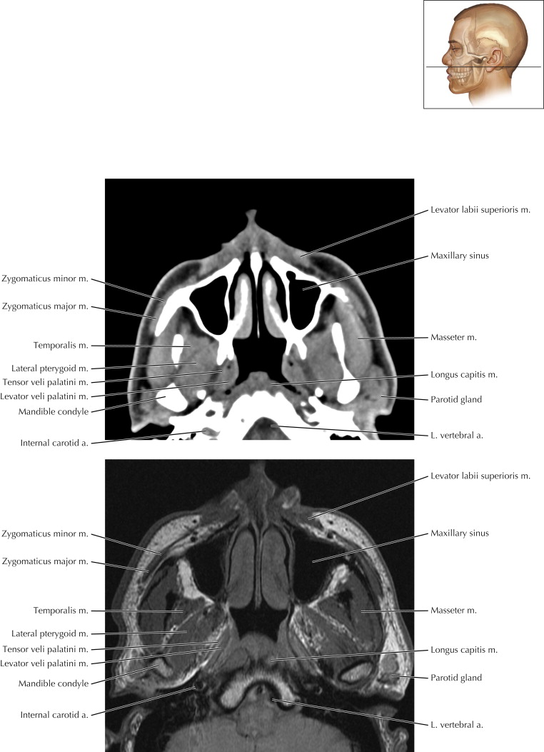
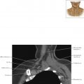
Four paired muscles—temporalis, masseter, medial pterygoid, and lateral pterygoid—are the primary muscles of mastication, responsible for adduction and lateral motion. In the axial MR image on the next page, note the temporalis muscle superficial to the temporal lobe of the brain.
These muscles are innervated by the mandibular branch (V 3 ) of the trigeminal nerve, cranial nerve (CN) V (see Axial 2). The lateral pterygoid helps open the mouth, and the medial pterygoid helps close the mouth (“lateral lowers and medial munches”).

Mandible and Muscles of Mastication Axial 2

Normal Anatomy
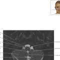
Note that from superficial to deep, the muscles are zygomaticus major, masseter, temporalis, and lateral pterygoid. The medial pterygoid muscle is located deeper and more caudal.
Pathologic Process
Denervation of the V 3 branch of the trigeminal nerve can lead to atrophy of the ipsilateral muscles of mastication.


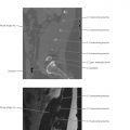
Mandible and Muscles of Mastication Axial 3