50% contain ectopic gastric mucosa

90% present with GI bleeding in children
IMAGING
•
Rule of 2s

Seen in ∼ 2% of population

Located within 2 feet of ileocecal valve

Length of 2 inches (on average)

Symptomatic usually before age 2

2 main complications in adults: Diverticulitis (20%) and intestinal obstruction (40%)
•
CT: Meckel diverticulitis

Blind-ending pouch containing fluid, air, or particulate matter (including calculi)

Inflamed: Mural thickening of diverticulum and adjacent SB

Shows mural enhancement on CECT

Mesenteric fat infiltration and fluid
–
Extraluminal gas and lymphadenopathy in some cases

± partial or complete small bowel obstruction

± intussusception
–
Inverted diverticulum may form lead mass
TOP DIFFERENTIAL DIAGNOSES
•
Mesenteric adenitis and enteritis
CLINICAL ISSUES
•
Children: Present with GI bleeding before age 2
•
Adults: Present with diverticulitis or obstruction
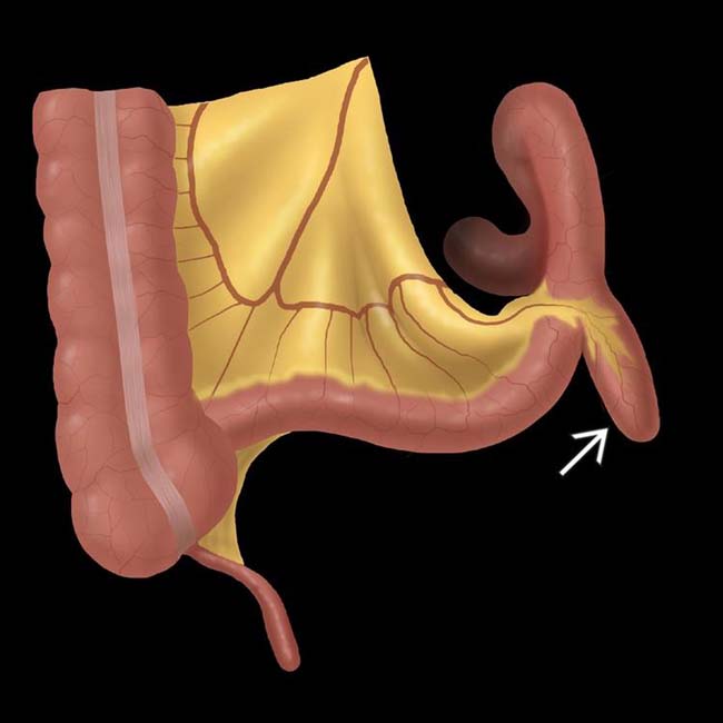
(Left) Graphic shows a blind-ended outpouching  from the antimesenteric border of the distal ileum, typical of a Meckel diverticulum.
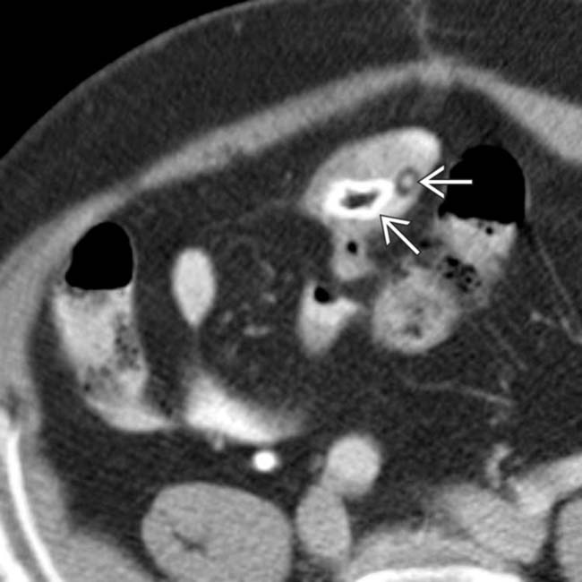
(Right) Axial CECT shows enteroliths  within a blind-ended sac in the right lower quadrant that proved to be a Meckel diverticulum at surgery.
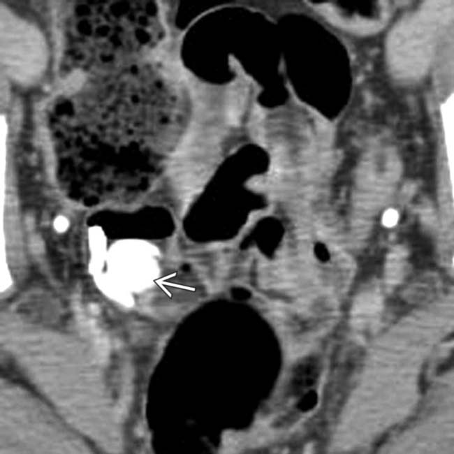
(Left) Axial CECT in a 27-year-old man with RLQ pain shows calcified enteroliths  lying within a blind-ending sac in the RLQ.
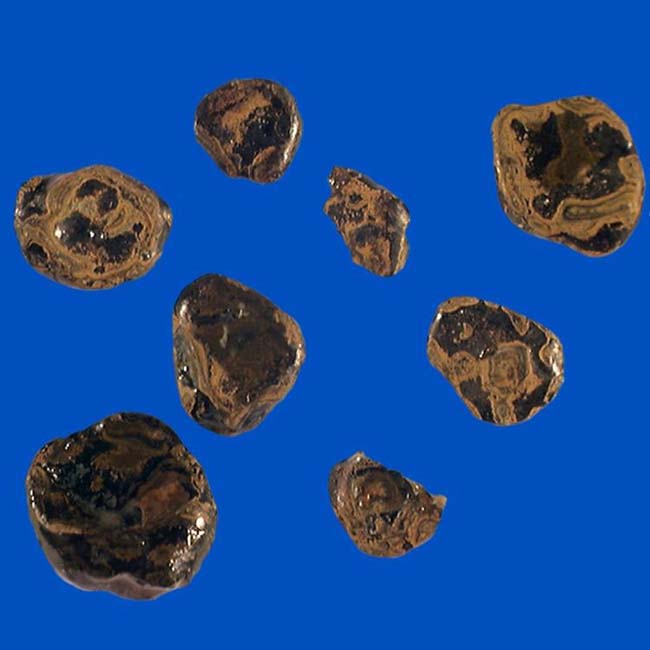
(Right) Surgery confirmed a Meckel diverticulum with mild chronic inflammation of the wall and multiple calcified enteroliths (shown here) within it.
TERMINOLOGY
Abbreviations
•
Meckel diverticulum (MD)
Definitions
•
Ileal outpouching due to persistence of omphalomesenteric or vitelline duct
IMAGING
General Features
•
Best diagnostic clue

Blind-ended sac or outpouching on antimesenteric border of distal ileum
•
Size

4-10 cm in length
•
Morphology

Tubular outpouching of ileum
•
Other general features

Most common congenital anomaly of GI tract

True diverticulum (contains all layers of bowel wall)

Arises from antimesenteric border of distal ileum

Formed by incomplete obliteration of ileal end of vitelline duct

Usually located within 50-60 cm of ileocecal valve

50% contain ectopic gastric mucosa
–
± pancreatic, duodenal, and colonic mucosa

90% of cases with bleeding contain gastric mucosa

Fibrous band (obliterated part of vitelline duct may connect apex of diverticulum to umbilicus)

Rule of 2s
–
Seen in ∼ 2% of population
–
Located within 2 feet of ileocecal valve
–
Length of 2 inches (on average)
–
Symptomatic usually before age 2
–
2 main complications in adults: Diverticulitis (20%) and intestinal obstruction (40%)
Radiographic Findings
•
Radiography

Radiograph A-P abdomen
–
Round collection of gas ± solitary or multiple calcified densities (enteroliths) within it in right lower quadrant (RLQ)
•
Fluoroscopic-guided enteroclysis

Superior due to maximum luminal distention

Blind-ended sac on antimesenteric border of ileum with either broad base or narrow neck

Broad-based diverticulum
–
Enteroclysis shows distinctive triangular junctional fold pattern at site of origin

Narrow neck diverticulum
–
Diagnosis depends on demonstration of blind end of diverticulum and its antimesenteric origin

Appears small initially but fills more completely with increased distention of lumen

Inverted Meckel diverticulum
–
Solitary, elongated, smoothly marginated, often club-shaped intraluminal mass parallel to long axis of distal ileum; may lead to intussusception
•
Double-contrast barium enema

Occasionally demonstrates MD by reflux into ileum


