and Vincent L. Sorrell2
(1)
Division of Nuclear Medicine and Molecular Imaging Department of Radiology, University of Kentucky, Lexington, Kentucky, USA
(2)
Division of Cardiovascular Medicine Department of Internal Medicine Gill Heart Institute, University of Kentucky, Lexington, Kentucky, USA
Electronic supplementary material
The online version of this chapter (doi: 10.1007/978-3-319-25436-4_11) contains supplementary material, which is available to authorized users.
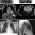
Table 11.1 outlines benign and malignant conditions affecting the mediastinum that may be detected during review of MPI projection data (Seo et al. 2005). The most common in routine clinical practice is hiatal hernia. Hiatal hernias are created when a portion of the stomach invaginates through the esophageal hiatal opening in the diaphragm and transiently or permanently resides above the diaphragm. Hiatal hernias can be quite large, and in those cases, most of the stomach becomes intrathoracic in location (Fig. 11.1). On SPECT MPI, hiatal hernias may appear “hot” when containing refluxed bile or “cold” when containing nonradioactive fluid or food (Burrell and MacDonald 2006; Chamarthy and Travin 2010; Gedik et al. 2007; Raza et al. 2005a; Slavin et al. 1998). “Hot” or “cold” hiatal hernias can be problematic because processing artifacts commonly lead to misinterpretation; thus, recognition of this common condition may be critical for proper reporting. Correlation with other imaging and clinical history is often essential.


Table 11.1
Differential diagnosis of “hot” and “cold” imaging findings related to the mediastinum
Organ system | “Hot” finding | “Cold” finding | References |
|---|---|---|---|
Mediastinum | Gastroesophageal reflux Hiatal hernia (refluxed duodenogastric bile) Neoplasm, primary malignant (esophageal cancer, lymphoma, thymoma) Extramedullary hematopoiesis (paravertebral masses) | Cyst Dilated esophagus fluid- or food-filled hiatal hernia | Burrell and MacDonald (2006) Chadika et al. (2005) Chamarthy and Travin (2010) García-Talavera et al. (2013) Gedik et al. (2007) Hawkins et al. (2007) Niederkohr et al. (2009) Raza et al. (2005a) Seo et al. (2005) Slavin et al. (1998) Vijayakumar et al. (2005) Watanabe et al. (1997) |


Fig. 11.1


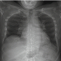
Huge hiatal hernia/intrathoracic stomach. In the thorax, there is a striking abnormality: lateral to the right side of the heart and behind the left side of the heart, there are large mixed “hot” and “cold” regions (a–d). Two selected contrast-adjusted stress images (c, d) orient the viewer to the locations of the abnormalities to best advantage. Re-reviewing the rest and stress raw images (a, b) with attention to these regions facilitates perception of the swirling radioactivity, suggesting duodenogastric reflux into the large herniated stomach. By radiography, there is a corresponding huge hiatal hernia/intrathoracic stomach behind and to the right of the heart (e, f). Note the normal thyroid gland at the top of the field-of-view on the rest raw images (a). MPI (g, h) shows a small-to-medium-sized, moderately severe, fixed anteroseptal-apical defect. There is normal LV function, regional wall motion, and wall thickening. The perfusion defect is likely artifactual in nature. LVEF is 64 %. RV function is normal (h).(a) Rest raw projection images (Video 11.1a, frame 1), at usual contrast setting for the heart, 99mTc sestamibi. (b) Stress raw projection images (Video 11.1b, frame 1) contrast-adjusted for intrathoracic abnormality, 99mTc sestamibi. (c) Stress raw projection image (Video 11.1c, frame 8), 99mTc sestamibi, activity lateral to the right heart (pink lines). (d) Stress raw projection image (Video 11.1c, frame 46), 99mTc sestamibi, activity behind the left heart (blue lines). (e) PA chest radiograph, right lateral margin of hiatal hernia (blue lines). (f) Lateral chest radiograph, approximate position of hiatal hernia containing gas (blue oval). (g) Stress/rest processed SPECT images (SA, HLA, VLA). (h) Stress gated SPECT images (Video 11.1d, frame 1) (SA, HLA, VLA)
Stay updated, free articles. Join our Telegram channel

Full access? Get Clinical Tree


