(1)
Department of Radiology, Saitama Medical University, Moroyama, Saitama, Japan
Abstract
Menisci are thought to play a role in maintaining the stability of the tibiofemoral joint, spreading the mechanical weight, shock absorption, and protection of cartilage. It is thought that menisci transmit 60–70% of mechanical weight to the femur and tibia.
7.1 Anatomy
Menisci are thought to play a role in maintaining the stability of the tibiofemoral joint, spreading the mechanical weight, shock absorption, and protection of cartilage. It is thought that menisci transmit 60–70% of mechanical weight to the femur and tibia.
Menisci are comprised of fibrous cartilage, mainly composed of type I collagen. The inner two third contains fibers running in the longitudinal and circumferential fashion. The outer one third is mainly composed of fibers running in the circumferential fashion. In the central (deep) portion of the meniscus, fibers run in a random manner.
The outer one third has vascular supplies and is called the red zone. If a small tear occurs here, natural healing may occur. However, the inner portion has no vascular supply and is called the white zone (Fig. 7.1).
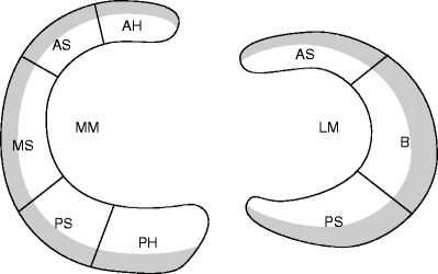
Fig. 7.1
Schematic illustration of the menisci. Medial meniscus (MM) and lateral meniscus (LM), which looks more like a C-shape. AH anterior horn, AS anterior segment, MS middle segment, PS posterior segment, PH posterior horn. The outer one third (shaded gray area) has vascular supplies and is called the red zone
There are two ways of classifying meniscal segments.
It can be divided into three segments: anterior segment, body, and posterior segment (or anterior horn, body, and posterior horn).
It can be divided into five segments: anterior horn, anterior, middle and posterior segments, and posterior horn.
7.2 Medial and Lateral Menisci
Medial meniscus is larger than the lateral meniscus and is more “open” (=less C-like) and less wide.
Peripheral part of the medial meniscus is thick, especially the posterior segment is the thickest at 5 mm approximately.
Posterior horn of the medial meniscus is always wider than the anterior horn and can be as wide as 12 mm (which is twice as wide as the anterior horn). The width of the posterior horn is greater than its thickness (Fig. 7.2).
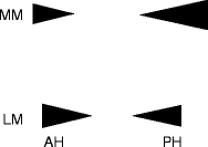
Fig. 7.2
Appearance of the medial (MM) and the lateral (LM) menisci in sagittal MRI. The posterior horn of the medial meniscus is wider and thicker than the anterior horn. This discrepancy is not seen in the lateral meniscus
Lateral meniscus has a smaller radius than the medial meniscus, and it has a more C-like shape.
Width of the lateral meniscus is consistently 10 mm approximately (Fig. 7.2).
Posterior horn of the lateral meniscus lies obliquely in the coronal MRI due to the slope of the tibial plateau. For this reason, it may be affected by the magic angle effect (see Chap. 2).
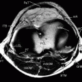
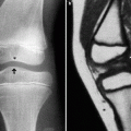
The tip of the anterior and posterior horns of the menisci are called “meniscal root.” They firmly attach to the tibia (Figs. 7.3 and 7.4). Posteriorly, the posterior root covers the surface of the tibial plateau.
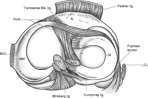
Fig. 7.3
Schematic illustration of the menisci and the surrounding structures sitting on the tibial plateau, as seen from above
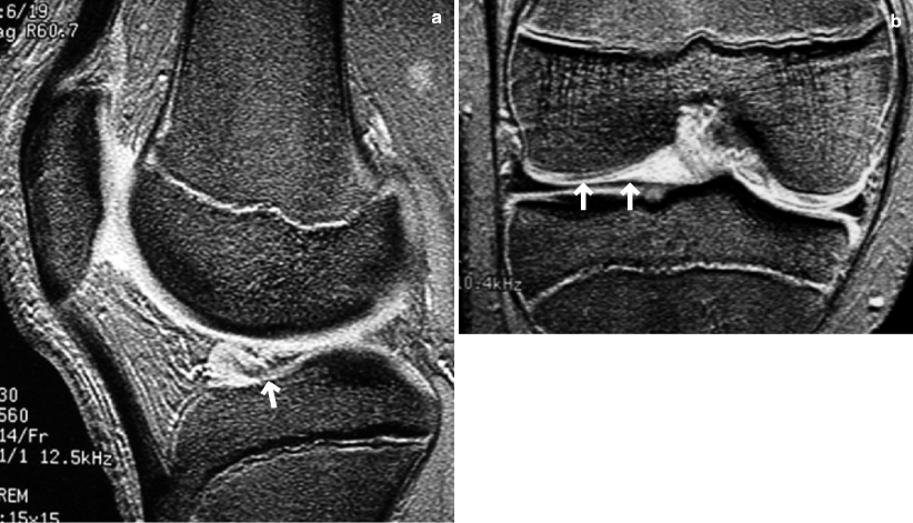
Fig. 7.4
Anterior meniscal root of the lateral meniscus. (a) Sagittal and (b) coronal T2*WI shows normal anterior meniscal root (arrows). It can be mistaken as a meniscal tear in a single sagittal slice, but the coronal image confirms it is a normal root
Anterior root of the medial meniscus attaches immediately anterior to the tibial ACL attachment site, while that of the lateral meniscus attaches immediately posterior to the tibial ACL attachment site.
Transverse ligament of Winslow connects between the medial and lateral menisci, both anteriorly (between anterior horns) and posteriorly (between posterior horns).
Posterior component of the anterior root of the medial meniscus joins the transverse meniscal ligament.
Peripheral part of the medial meniscus firmly attaches to the deep layer of the MCL and the joint capsule.
Lateral meniscus has a weak bond with the joint capsule, and posteriorly it is only supported by the limited amount of fascicles of the joint capsule. Thus, its range of motion is much greater than that of the medial meniscus as the knee is flexed and extended.
7.3 Delineation of Meniscal Lesions by MRI
High-resolution MRI is essential for delineation of a small meniscal tear.
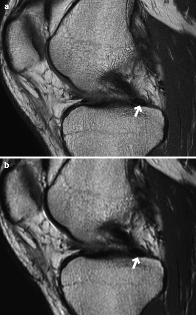
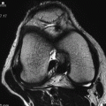
Fig. 7.5
Posterior meniscal root of the medial meniscus. PDWI showing the posterior root of the medial meniscus. (a) 512 sampling and 1024 matrix and (b) 256 sampling and 256 matrix. High-resolution image (a) enables visualization of the fibers of the posterior root of the medial meniscus (arrows)
Meniscal window
Delineation of meniscal lesions requires the use of “meniscal window,” in which the window width is kept narrow and the window level low (Fig. 7.6). Therefore, if the images need to be printed onto a film, two sets of films with different window levels (one for visualization of meniscal lesions and the other for other structures of the knee) may be needed.
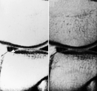
Fig. 7.6
Meniscal window. “Meniscal window” (left) with a narrow window width and a low window level, and (right) normal window width/level. PDWI. A small lesion in the anterior segment of the lateral meniscus (arrow) can only be detected in the “meniscal window” image. Note, however, it is impossible to see anything within the bone if the “meniscal window” is used
FSE vs. conventional SE
When the fast spin-echo (FSE) sequence initially became available, its ability to delineate meniscal lesions was reported to be inferior to that of conventional SE sequence (Rubin et al. 1994).
However, later reports emphasized the usefulness of proton-density-weighted and T2-weighted FSE sequences (Escobedo et al. 1996).
FSE sequence with echo train length of 5–6 produces images without blurring, and its short scan time allows for higher spatial resolution and increased NEX.
References
Rubin DA, Kneeland JB, Listerud J, Underberg-Davis SJ, Dalinka MK. MR diagnosis of meniscal tears of the knee: value of fast spin-echo vs conventional spin-echo sequences. AJR. 1994;162:1131–5.
Escobedo EM, Hunter JC, Zink-Brody GC, Wilson AJ, Harrison SD, Fisher DJ. Usefulness of turbo spin-echo MR imaging in the evaluation of meniscal tears: comparison with a conventional spin echo sequence. AJR. 1996;167:1223–7.
7.4 Meniscal Tear
Definition of meniscal tear
1.
Intrasubstance hyperintensity that reaches the surface of the meniscus
2.
Deformation of the meniscus
The posterior segment of the medial meniscus is the thickest portion of the medial meniscus, and light intrasubstance hyperintensity is often observed. However, unless the hyperintensity reaches the meniscal surface, it is more likely to be physiologic changes (mucoid or myxoid degeneration) (Figs. 7.7 and 7.8).
There is no evidence to suggest that intrasubstance hyperintensity within the meniscus seen in young persons will develop into a meniscal tear at later life.
If signal changes suggestive of a meniscal tear are seen in more than one slices, or both in sagittal and coronal slices, the signal change is likely to represent a true meniscal tear (sensitivity >90%).
If such signal changes are seen in only one slice, the sensitivity is 55% for the medial meniscus and 30% in the lateral meniscus.

Fig. 7.7
Meniscal intrasubstance hyperintensity (degeneration)
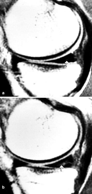
Fig. 7.8
Intrasubstance hyperintensity within the posterior segment of the medial meniscus. PDWI. Within the posterior segment of the medial meniscus, there is dotted (a) and linear (b) hyperintensity (arrows). If such signal changes do not reach the meniscal surface, it is more likely to be physiologic degenerative changes rather than a tear
Reference
De Smet AA, Norris MA, Yandow DR, et al. MR diagnosis of meniscal tears of the knee: importance of high signal in the meniscus that extends to the surfact. AJR. 1993;161:101–7.
Classification of the meniscal tear
1.
Vertical tear
(a)
Longitudinal tear
Bucket-handle tear
(b)
Radial tear
(c)
Oblique tear (parrot-beak tear)
2.
Horizontal tear
3.
Complex tear
(A)
Complete tear (full-thickness tear)
(B)
Partial tear
If the tear reaches both the superior and the inferior meniscal surface, it is called a complete tear (or a full-thickness tear). If the tear reaches only one surface, it is called a partial tear.
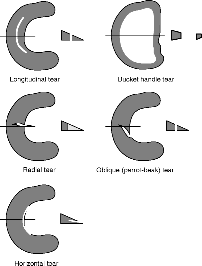
Fig. 7.9
Classification of meniscal tear (1)
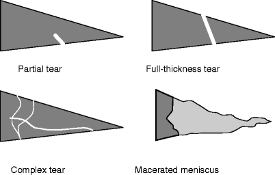
Fig. 7.10
Classification of meniscal tear (2)
Vertical tear
Vertical tear is commonly seen in young persons.
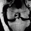
Longitudinal tear commonly begins in the posterior horn and extends into the middle segment (Fig. 7.11).
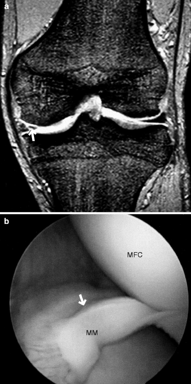
Fig. 7.11
Longitudinal tear of the posterior horn. A man in his 20s. (a) Coronal T2*WI and (b) arthroscopic image. There is a full-thickness longitudinal tear extending from the posterior horn to the middle segment of the medial meniscus (MM) (arrows). MFC medial femoral condyle
In sagittal image, vertical tear may be partially unclear due to partial volume effect.
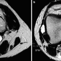
Radial tear usually begins from the free border of the meniscus and extends into the peripheral portion.
Radial tear may be difficult to visualize, especially in the axial slice.
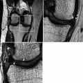
Radial tear sometimes produces a meniscal gap, depending on the slice (Fig. 7.12).
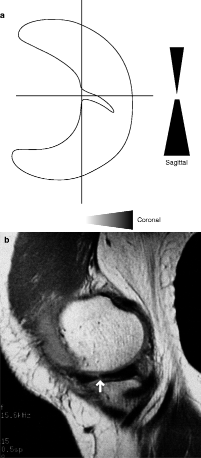
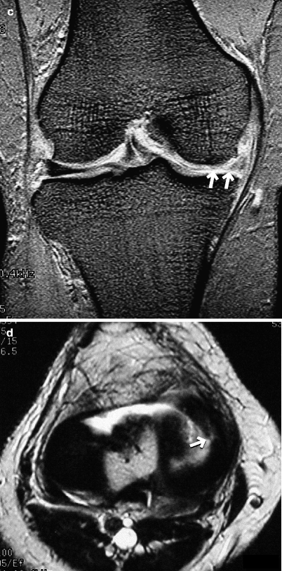
Fig. 7.12
Radial tear. A woman in her 50s. (a) Schematic illustration, (b) PDWI, (c) coronal T2*WI, and (d) axial T2WI. There is a full-thickness tear extending from the free border of the middle segment to the peripheral direction (arrow, b). In the coronal slice which cuts through the radial tear, the meniscus partially disappears (arrows, c). It is rare for a radial tear to be entirely visualized in axial images (arrow, d)
Radial tear of the meniscal root is particularly difficult to diagnose.
If radial tear progresses, a portion on the side of free border becomes detached from the meniscal body and creates a flap (Fig. 7.13).
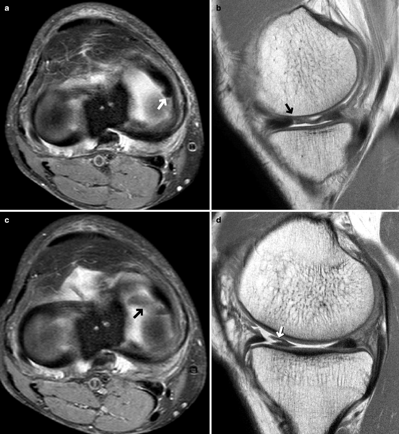
Fig. 7.13
Radial tear which progressed to a flap tear. A woman in her 50s. (a, b) Taken at the time of initial presentation. (c, d) Taken at 40-day follow-up. (a, c) Axial FS PDWI. Initially, there was only a radial tear of the middle segment of the medial meniscus (a, b, arrows) but at follow-up, the torn fragment is anteriorly displaced to form a flap (c, d, arrows)
A fragment from the flap tear may drop into the coronary recess of the joint space (Fig. 7.14).
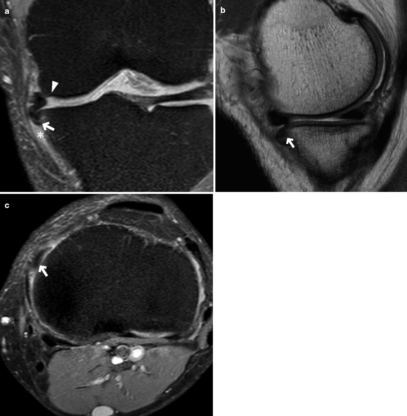
Fig. 7.14
Fragmentof a flap tear that fell into the coronary recess. A man in his 30s. (a) Coronal FS PDWI, (b) PDWI, and (c) axial FS PDWI. A fragment (arrow) of a flap tear (arrowhead, a) is seen falling into the coronary recess (*, a) of the joint space
If a fragment is displaced to the intercondylar space (especially the lateral meniscus), it will be positioned just inferoposterior to the ACL, leading to the “double-ACL” sign (there appears to be the “second ACL” under the true ACL) (Fig. 7.15).
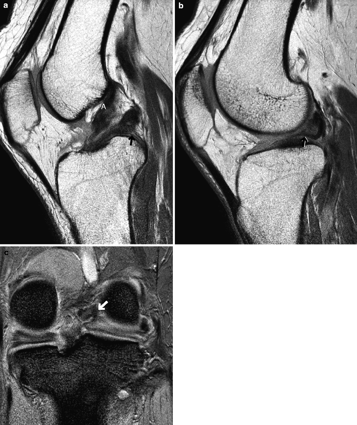
Fig. 7.15
Double ACL sign created by a fragment of the lateral meniscal flap tear. A woman in her 50s. (a, b) PDWI and (c) coronal T2*WI. Slice (b) is more lateral to the slice (a). A fragment of the flap tear (arrows) is displaced to the intercondylar space, and there appears to be the “second ACL” just inferoposterior to the true ACL (A)
References
Lecas LK, Helms CA, Kosarek FJ, Garret WE. Inferiorly displaced flat tears of the medial meniscus: MR appearance and clinical significance. AJR. 2000;174:161–4.
Vande Berg BC, Malghem J, Pilvache P, maldague B, Lecouvet FE. Meniscal tears with fragments displaced in notch and recesses of knee: MR imaging with arthroscopic comparison. Radiology. 2005;234:842–50.
Bui-Mansfield LT, Dewitt RM. Magnetic resonance imaging appearance of a double anterior cruciate ligament associated with a displaced tear of the lateral meniscus. J Comput Assist Tomogr. 2006;30:327–32.
Horizontal tear
Horizontal tear is commonly seen in elderly persons and frequently coexists with intrasubstance signal abnormalities (Fig. 7.16). It is also called “fish mouth” or “cleavage.”
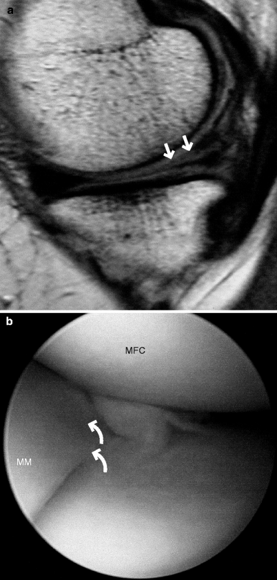
Fig. 7.16
Horizontal tear. A woman in her 50s. (a) PDWI and (b) arthroscopic image. There is a horizontal tear extending from the free border of the meniscus within the posterior segment of the medial meniscus (arrows, a). Arthroscopy revealed a horizontal tear (curved arrows, b) in the middle and posterior segments of the medial meniscus
Horizontal tear begins just below the meniscal surface and extends peripherally.
If the tear reaches the peripheral border, joint fluid will enter through it to form a meniscal cyst (see Chap. 12).
Complex tear
Complex tear is the most severe form of tear and involves both vertical and horizontal tears. It can develop into meniscal segmentation and displacement of the entire meniscus from its original position (Fig. 7.17).
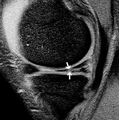
Fig. 7.17
Complex tear. A man in his 20s. Sagittal T2*WI. There are several tears (arrows) in addition to a horizontal tear within the posterior segment of the medial meniscus
Complex tear and severe degenerative changes will lead to macerated meniscus, which is the end stage of the meniscal damage (Fig. 7.18).
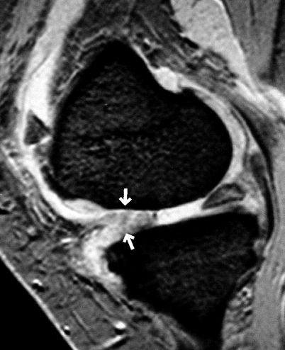
Fig. 7.18
Maceratedmeniscus. A man in her 60s. Sagittal T2*WI. The anterior segment of the medial meniscus has lost its original shape and appears swollen (arrows) due to complex tear and severe degenerative changes
Meniscal contusion
Localized intrasubstance hyperintensity following acute meniscal trauma (Fig. 7.19).Get Clinical Tree app for offline access
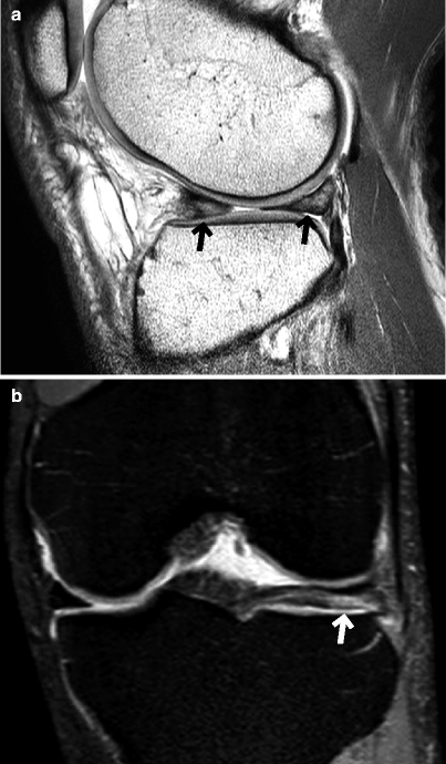
Fig. 7.19
Meniscal contusion. A man in his 20s who had a fall from a height 1 month prior to presentation. (a) PDWI and (bRelated posts:

Stay updated, free articles. Join our Telegram channel

Full access? Get Clinical Tree