(1)
Department of Radiology, Saitama Medical University, Moroyama, Saitama, Japan
Abstract
Layer I: Thin sheet that overlies the two heads of the gastrocnemius and the structures of the popliteal fossa.
5.1 Anatomy
MCL generally consists of three layers (Figs. 5.1 and 5.2), but the naming and classification of the layers that comprise MCL may vary according to different authors:
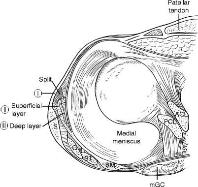
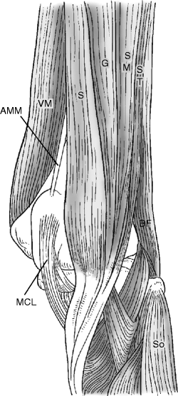
Fig. 5.1
Three layers of the MCL. Layer I: thin sheet that overlies the two heads of the gastrocnemius and the structures of the popliteal fossa. Layer II: superficial layer of MCL. Layer III: medial joint capsule including the deep layer of MCL. S sartorius, G gracilis, ST semitendinosus, SM semimembranosus, mGC medial head of gastrocnemius (Illustration adapted from Warren and Marshall)
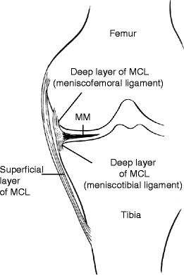
Fig. 5.2
Schematic illustration of the MCL. The superficial layer (also known as the tibial collateral ligament) and the deep layer (also known as medial capsular ligament). Superficial layer of MCL attaches to the tibia at 7–8 cm below the joint space (note the significant distance). The deep layer firmly attaches to the medial meniscus and is also known as meniscofemoral and meniscotibial ligament
Layer I: Thin sheet that overlies the two heads of the gastrocnemius and the structures of the popliteal fossa.
Layer II: Superficial layer of the MCL (alternatively called tibial collateral ligament). Anteriorly, Layer II blends with Layer I through the split to form the medial patellar retinaculum. Posteriorly, it blends with Layer III via the posterior oblique ligament.
Layer III: Deepest layer of the MCL called medial capsular ligament, which is continuous with the medial joint capsule.
Fibrofatty tissue fills the space between Layer I and II, and the tendons of semitendonisus and gracilis run through this space.
Small bursae are located within fibrofatty tissue between Layer II and II (Fig. 5.3).
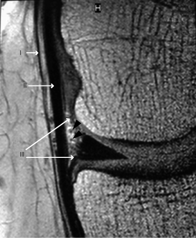
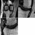
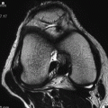
Fig. 5.3
High-resolution image of the medial compartment of the knee joint acquired using a microscopy coil (FOV 50 mm, slice thickness 1.5 mm). As shown in Fig. 5.1, layer I: thin sheet of fascia, layer II: superficial layer of the medial collateral ligament (MCL), layer III: deep layer of MCL, and a small bursa and small blood vessels (arrowheads)
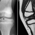
Superficial layer of MCL runs vertically and has a width of 15 mm, length of 8–12 cm, and thickness of 2–3 mm.
The posterior oblique portion of the MCL (posterior oblique ligament) is fused with layer III and closely attached to the medial meniscus and also the tibia (Fig. 5.4).
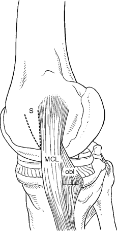
Fig. 5.4
Superficiallayer of the medial collateral ligament (MCL) and posterior oblique ligament (obl). There is a split (S) anterior to the superficial layer of the MCL (see also Fig. 5.1)
Superficial layer of MCL proximally attaches to the medial femoral condyle 5 cm above the joint space and distally attaches to the metaphyseal region of the tibia 6–7 cm below the joint space. For this reason, on MR imaging, care must be taken to include the inferior edge of the distal MCL within the FOV. The distal attachment lies beneath the pes anserinus.
Deep to the vertical component of the superficial MCL, the capsule becomes thicker, forming the deep layer of MCL. This layer inserts directly into the edge of femur and tibial plateau and firmly attaches to the medial meniscus and thus divided into meniscofemoral and meniscotibial ligaments, respectively. However, in normal knees without joint effusion, these ligaments may not be delineated on MRI (Figs. 5.5).
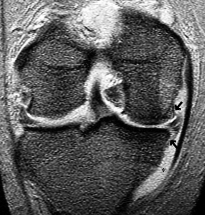
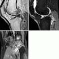
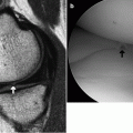
Fig. 5.5
Visualization of the deep layer of MCL (arrow) due to the presence of joint effusion. Damage to MCL or the medial meniscus led to accumulation of joint fluid between the superficial and deep layers of the MCL (*), enabling the delineation of the deep layer