(1)
Department of Radiology, Saitama Medical University, Moroyama, Saitama, Japan
Abstract
The lateral supporting structure of the knee is a compound of several ligaments and tendons and thus is more complex than the medial supporting structure.
6.1 Anatomy
The lateral supporting structure of the knee is a compound of several ligaments and tendons and thus is more complex than the medial supporting structure.
It consists of three layers:
Layer I: Has two parts: the iliotibial band (ITB) and its expansion anteriorly and the superficial portion of the biceps femoris tendon (BFT) and its expansion posteriorly.
Layer II: Anteriorly it is formed by the lateral patellar retinaculum, and laterally by the lateral collateral ligament (LCL, also known as fibular collateral ligament).
Layer III: This is the deepest layer which is the lateral part of the joint capsule and includes fabellofibular ligament and arcuate ligament.
Three most prominent structures are, from anteriorly, ITB, LCL, and BFT (Fig. 6.1).
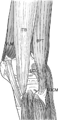
Fig. 6.1
Schematicillustration of the lateral supporting structures. From anteriorly, iliotibial band (ITB), lateral collateral ligament (LCL), and biceps femoris tendon (BFT). VLM Vastus lateralis, IGCM Lateral head of gastrocnemius
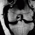
LCL runs down to form a conjoint tendon with BFT near the attachment site of the fibular head, where it forms a V-shape. However, BFT belongs to the Layer I and the LCL belongs to the Layer II, anatomically and it is uncommon to be able to visualize this V-shape in a single sagittal image(Fig. 6.2).
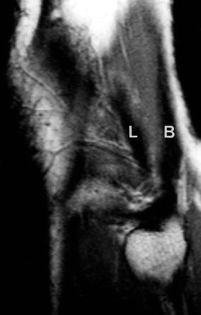
Fig. 6.2
Conjoint tendon. This PDWI shows LCL (L) and BFT (B) form the conjoint tendon. Anatomically it forms a V-shape. However, it is uncommon to be able to visualize this V-shape in a single sagittal image
LCL resists the varus stress and prevents overextension of the knee. It is under the highest tension when the knee is extended.
Popliteus tendon runs in a slightly different route than the aforementioned three ligaments (Fig. 6.3).
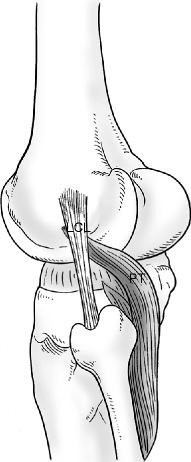
Fig. 6.3
Schematic illustration of the lateral collateral ligament and the popliteus tendon. Popliteus tendon (PT) arises from the lateral femoral condyle and runs deep to the LCL through the popliteal hiatus to reach the extra-articular space
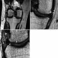
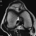
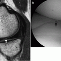
Popliteus tendon arises from the popliteal sulcus (Fig. 6.4) located just below the LCL attachment site of the lateral femoral condyle. It runs deep to the LCL, passes diagonally through the meniscocapsular junction of the lateral meniscus to become the popliteus muscle, and then attaches to the posterior surface of the tibia. Thus, the upper half of the popliteal tendon is intra-articular and can be observed by arthroscopy (Fig. 6.5). One should be cautious not to misdiagnose the normal popliteal tendon sheath which runs close to the periphery of the lateral meniscus as a meniscal tear (Figs. 6.6 and 6.7).
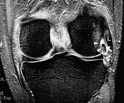
Fig. 6.4
Poplitealsulcus of the lateral femoral condyle. Coronal T2*WI shows the popliteus tendon (arrow) arising from the popliteal sulcus (arrowheads), running deep to the LCL (L)
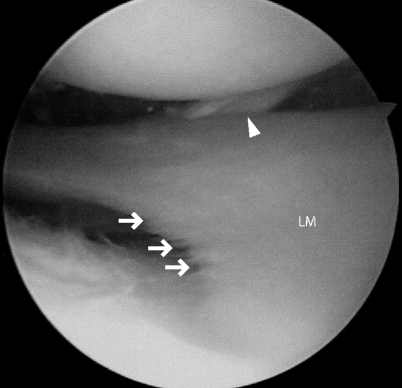
Fig. 6.5
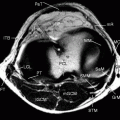
Popliteus tendon as seen by arthroscopy. Popliteus tendon is seen running diagonally beyond the lateral meniscus (LM). Free border of the meniscus shows fibrillation as a result of degenerative process
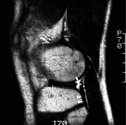
Fig. 6.6
Popliteus tendon. T2WI through the lateral meniscus (LM). Popliteus tendon (arrowheads) forms tendon sheath and runs through the meniscocapsular junction of the lateral meniscus. It is physiologic to see some fluid accumulation (arrow) at this location, and this should not be mistaken as meniscal damage
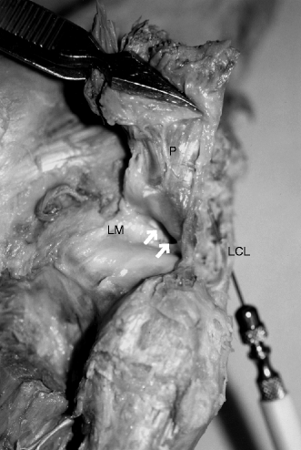
Fig. 6.7
Cadaveric specimen of popliteus tendon, LCL and lateral meniscus. Popliteus tendon (P) is cut through the musculotendinous junction and folded over to reveal the space (arrows) between it and the lateral meniscus (LM). The needle is placed deep to the LCL
MRI may reveal a thin band-like structure called popliteofibular ligament which arises from the popliteus tendon and inserts to the fibular head (Figs. 6.8 and 6.9).
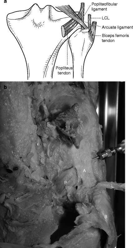
Fig. 6.8
Schematic illustration (a) and cadaveric specimen (b) of the posterolateral corner of the knee. Popliteus tendon (P), which gives off a small branch called popliteofibular ligament that attaches to the fibular head. The musculotendinous junction of the popliteus muscle runs deep to the arcuate ligament (A) and reaches the popliteal fossa. L: LCL, B: BFT
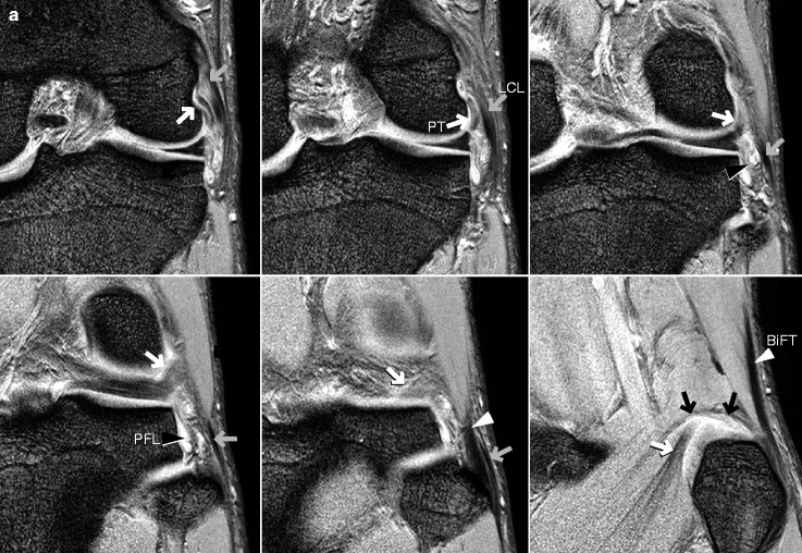
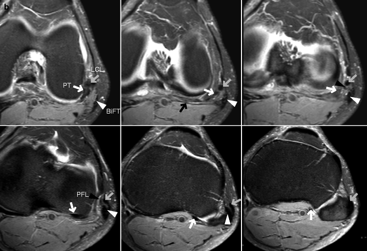
Fig. 6.9
MRI of the posterior structures of the knee. (a) Coronal T2*WI and (b) axial FS PDWI. Popliteus tendon (PT, white arrows) originates from just below the LCL (gray arrows) attachment site of the lateral femoral condyle, runs inferiorly, and gives off a branch called popliteofibular ligament (PFL, black arrowheads). The popliteus musculotendinous junction runs beneath the arcuate ligament (black arrows) and reaches the popliteal fossa. Attached to the fibular head are several ligaments and tendons including PFL, LCL, biceps femoris tendon (BiFT, white arrowheads)
Musculotendinous junction of the popliteus muscle is covered by the arcuate ligament (Fig. 6.10).Get Clinical Tree app for offline access
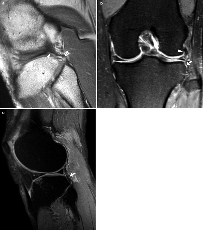
Fig. 6.10
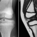
Damage to the popliteofibular ligament. (a) PDWI and (bRelated posts:

Stay updated, free articles. Join our Telegram channel

Full access? Get Clinical Tree