(1)
Department of Radiology, Saitama Medical University, Moroyama, Saitama, Japan
Abstract
Sagittal view is the basis for knee MRI including ACL evaluation. To improve delineation of ACL, the knee is slightly flexed (see Chap. 3 for more details). If the slice thickness is 3 mm or so, it is not possible to visualize ACL and PCL in their entire lengths in one plane. Menisci are depicted as bow-tie-shaped structure with homogeneous hypointensity.
1.1 Sagittal Views
3.0 mm slice thickness/0.3 mm interslice gap, 150 mm FOV, 512 × 256 matrix
(a)
Intermediate-weighted (close to proton density-weighted) FSE, TR/TE = 1,321/17 ms, ET = 5
(b)
T2*-weighted GRE, TR/TE = 522/14, flip angle 30°
Sagittal view is the basis for knee MRI including ACL evaluation. To improve delineation of ACL, the knee is slightly flexed (see Chap. 3 for more details). If the slice thickness is 3 mm or so, it is not possible to visualize ACL and PCL in their entire lengths in one plane. Menisci are depicted as bow-tie-shaped structure with homogeneous hypointensity.
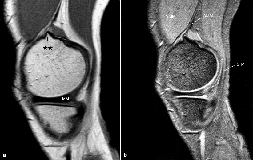
Fig. 1.1
(a) MM medial meniscus, ✩✩ adductor magnus muscle insertion, where distal femoral cortical irregularity may arise. (b) VMM vastus medialis muscle, GrM gracilis muscle, AMM adductor magnus muscle
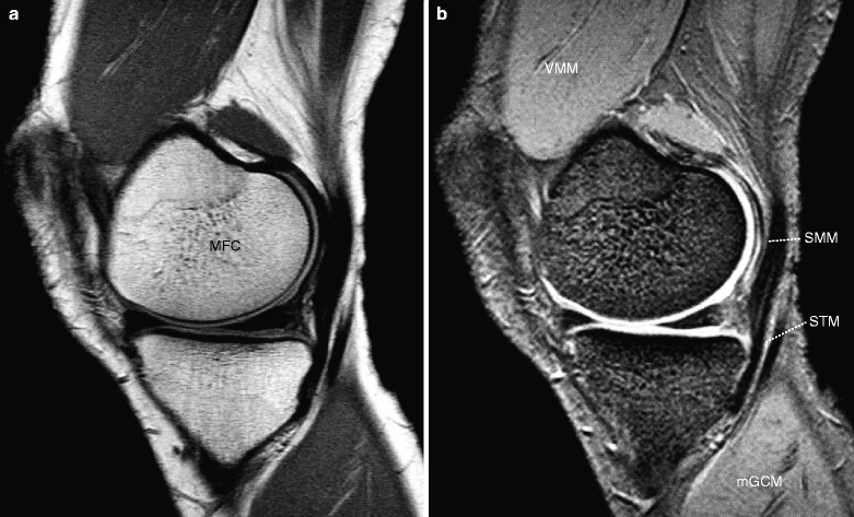
Fig. 1.2
(a) MFC medial femoral condyle. (b) VMM vastus medialis muscle, SMM semimembranosus muscle, STM semitendinosus muscle, mGCM medial head of gastrocnemius muscle
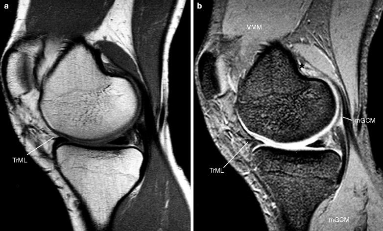
Fig. 1.3
(a) TrML transverse meniscal ligament. (b) mGCM medial head of gastrocnemius muscle, VMM vastus medialis muscle, TrML transverse meniscal ligament, ✩ distal femoral cortical irregularity at the insertion of mGCM
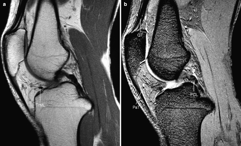
Fig. 1.4
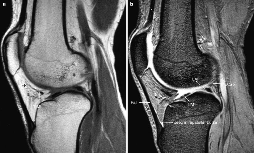
(a) iFP infrapatellar (Hoffa’s) fat pad, TrML transverse meniscal ligament. (b) QT quadriceps femoris tendon, PCL posterior cruciate ligament, PaT patellar tendon
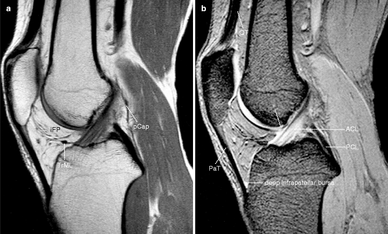
Fig. 1.5
(a) iFP infrapatellar (Hoffa’s) fat pad, pCap posterior capsule, TrML transverse meniscal ligament. (b) QT quadriceps femoris tendon, ACL anterior cruciate ligament, PCL posterior cruciate ligament, PaT patellar tendon, deep infrapatellar bursa, * location where infrapatellar plica and infrapatellar (Hoffa’s) fat pad curves in toward the femur