and Zdeněk Fryšák1
(1)
Department of Internal Medicine III – Nephrology, Rheumatology and Endocrinology, Faculty of Medicine and Dentistry, Palacky University Olomouc and University Hospital Olomouc, Olomouc, Czech Republic
Keywords
Thyroid carcinomaTotal thyroidectomyMetastatic cervical lymph nodesLocation of cervical lymph nodes19.1 Essential Facts
The risk factors influencing recurrence after the initial operation on PTC includes male sex, extrathyroid extension, metastatic lymph nodes (LN), distant metastasis, tumor size greater than 2 cm, subtotal thyroidectomy and without postoperative radioiodine 131I-therapy (RIT) [1].
Relapse of PTC can occur in three forms: distant metastasis, “true” local recurrence, and disease within metastatic lymph nodes. It is estimated that 90% of disease relapse in PTC are metastatic LNs. Because of the typically indolent nature of the disease, relapse is typically identified within the first 3–4 years [2].
Persistent or recurrent metastatic disease in patients with PTC detected during the follow-up period was found to be 20–28% [3].
In a study by Mazzaferri and Jhiang in the mid-1990s, post-treatment recurrence rates among PTC patients were 43.3% after more than 5 years of follow-up, and 19.3% more than 10 years after the original treatment [4].
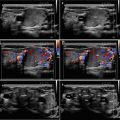
A later study by Durante et al. [2] reported that recurrences more than 5 years after surgery were discovered in 23% of patients (Figs. 19.6, 19.7 and 19.10). The differences between the findings of these two studies are largely a reflection of the changing demography of the differentiated thyroid carcinoma. Today’s PTCs are much more likely to be diagnosed at a subclinical stage than those treated in 1960s to the 1990s.
Follow-up of low risk patients with PTC remains questionable. Neck US, serum thyroglobulin (Tg), and whole-body scan after l-thyroxin withdrawal are performed.
Torlontano et al. reported that up to 50% of metastatic LNs were <1 cm and not palpable; negative predictive value of both negative Tg and US at first follow-up was 98.8% [5].
19.2 US Features of Metastatic Lymph Nodes
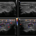
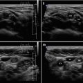
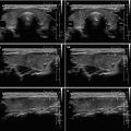
Metastatic LNs tend to be round (Figs. 19.2 and 19.8), hypoechoic (Figs. 19.1 and 19.9), and hyper vascularized (Fig. 19.6) with a loss of hilar architecture (all figures in this chapter), and in differentiated thyroid cancer—PTC, FTC (Fig. 19.2aa, bb) they may also demonstrate specific features such as hyperechoic punctuations (Figs. 19.3 and 19.4) or microcalcifications and cystic appearance (Figs. 19.5 and 19.7) [3].
LN round shape was reported to have an excellent specificity in 86%, but low sensitivity in 53% (likely partial or initial involvement of LN does not change the LN shape). In addition, some normal LNs may be rounded, especially in the parotid and submandibular regions [3].
Absence of echogenic hilum is a US sign with higher sensitivity of 92%, but with low specificity of 52% compared to LN round shape [6].
Presence of a hyperechoic hilum of the nodes is usually considered as a strong diagnostic criterion for benign LNs. However, it has been reported that 84–92% of benign nodes, but less than 5% of metastatic nodes, have a hyperechoic hilum [6].
A recent study compared roles of US, CT, and PET-CT in the evaluation of cervical recurrence in DTC, by correlating findings with sample pathology. Sensitivity, specificity, and accuracy were ≈69%, ≈90%, and ≈80% for US; ≈63%, ≈95%, and ≈80% for CT; and ≈54%, ≈79%, and ≈67% for PET-CT, respectively. Sensitivity and specificity of ultrasound and CT were higher than reported for PET-CT [7].
PET-CT is a valid imaging modality for the diagnosis of iodine-negative lesions on iodine scan [8].
Location of cervical lymph nodes—numerical classification system [9]:
C-IA—Submental lymph nodes.
C-IB—Submandibular lymph nodes.
C-II—Internal jugular (deep cervical) chain from the base of the skull to the inferior border of the hyoid bone.
C-III—Internal jugular (deep cervical) chain from the hyoid bone to the inferior border of the cricoid arch.
C-IV—Internal jugular (deep cervical) chain between the inferior border of the cricoid arch and the supraclavicular fossa.
C-V—Posterior triangle or spinal accessory nodes.
C-VI—Central compartment nodes from the hyoid bone to the suprasternal notch.
C-VII—Nodes inferior to the suprasternal notch in the upper mediastinum.

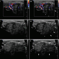
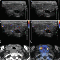
Fig. 19.1
(aa) A 39-year-old woman, 10 years post thyroidectomy for papillary thyroid carcinoma (PTC). Small solitary metastatic lymph node (LN) along the left internal jugular vein (IJV) at level C-IV, size 18 × 8 × 6 mm and volume 0.5 mL. US scans: ln (marks)—elliptical shape; homogeneous structure; hypoechoic; no hilus sign; transverse. (bb) Detail of solitary metastatic LN, CFDS: ln (marks)—hilar vascularity, one central vessel branch; transverse. (cc) Detail of solitary metastatic LN: ln (marks)—elliptical shape, size 18 × 8 mm, L/S ratio > 2 (not pathological); no hilus sign; longitudinal


