Miscellaneous Conditions
Thomas H. Berquist
Bone Islands (Enostosis)
Key Facts
Bone islands are benign sclerotic areas in bone. They may be single or multiple.
Bone islands typically are noted incidentally on radiographs.
Lesions may be seen in patients from 7 to 78 years of age. There is no sex predilection.
The most common sites are the ribs, pelvis, and femora. Up to 32% may change in size.
Radiographic features:
Round, oval, or spiculated sclerotic areas typically (66%) are 0.5 to 1.5 cm.
Appearance usually is characteristic, although the differential diagnosis could include blastic metastasis, osteoma, osteoid osteoma, or infarct.
Other imaging studies usually are not required. Radionuclide bone scans typically are normal, but focal increased tracer can occur.
Magnetic resonance imaging (MRI) shows low signal intensity on T1- and T2-weighted sequences.
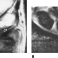
 FIGURE 15-1 Bone island in the femoral neck in a 60-year-old man. Anteroposterior (AP) (A) and oblique (B) radiographs demonstrate a spiculated sclerotic focus (arrow). Coronal T1-weighted (C), axial proton density (D), and T2-weighted (E) images showing a low-intensity lesion on all sequences with irregular margins (arrow). There is a small bone island in the opposite femoral head (open arrow) on the axial images (D,E). |
Suggested Reading
Hall FE, Goldberg RP, Davies JAK, et al. Scintigraphic assessment of bone islands. Radiology 1980;135:737–742.
Osteopoikilosis
Key Facts
Osteopoikilosis is a sclerotic bone dysplasia presenting in childhood. It has been detected in all bones except the skull.
Lesions are smaller than typical bone islands (2–10 mm).
The condition is considered an autosomal dominant chondrodysplasia.
Lesions may grow in children but stabilize or disappear in adults.
Most patients are asymptomatic, although 20% may present with joint pain.
Radiographic features:
Lesions are smaller and more well-defined than bone islands and involve the epiphysis and metaphysis.
Features are so characteristic that there usually is no difficulty in diagnosis.
Differential considerations include mastocytosis and tuberous sclerosis.
Radionuclide scans typically are normal but may be positive in growing lesions.
 FIGURE 15-2 Osteopoikilosis. AP radiograph of the pelvis and hips showing multiple small sclerotic foci in the proximal femora, ischia, and acetabuli. Note the femoral striations (arrows) (osteopathia striata is related to osteopoikilosis). |
Suggested Reading
Green AE, Ellowood WH, Collins JR. Melorheostosis and osteopoikilosis. AJR Am J Roentgenol 1962;87:1096–1117.
Osteopathia Striata
Key Facts
Osteopathia striata is a rare autosomal dominant inherited condition related to osteopoikilosis.
Patients usually are asymptomatic.
Radiographic features:
Distinct striations in the metaphysis of long bones parallel to the shaft. Striations may extend into the epiphysis.
Changes usually are bilateral.
The tibia is the most common site.
Radionuclide scans are normal.
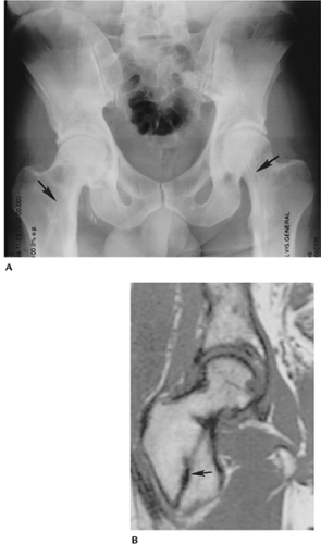 FIGURE 15-3 Osteopathia striata. (A) AP radiograph of the pelvis and hips showing linear striations in the femoral neck (arrows) with cortical thickening medially from associated melorheostosis. (B) Coronal T1-weighted magnetic resonance (MR) image of the right hip showing the low signal intensity linear sclerosis (arrow). |
Suggested Reading
Hurt RL. Osteopathia striata. Voorhoeve disease. J Bone Joint Surg 1953;35B:89–96.
Melorheostosis
Key Facts
Melorheostosis causes bone sclerosis involving one side of the cortex. The original description looked like dripping candle wax, thus the term “melorheostosis.”
Cause is unknown. Patients may be asymptomatic or present with pain in the involved region.
The condition may be present from birth to late adult life. In 50% of cases, the condition is evident by 20 years of age. There is no sex predilection.
The involved extremity may be shorter or, in some cases, longer. Muscle atrophy also is present in some cases.
Radiographic features:
The condition most commonly involves the long bones of the extremities. Most often it is unilateral.
Sclerosis and cortical thickening involve one side of the involved bone or bones. Typically, the process extends into the metaphysis or epiphysis, but soft tissue and joint involvement can occur. Associated conditions include:
Leg length discrepancy
Scleroderma
Neurofibromatosis
Osteopoikilosis
Osteopathia striata
Hemangiomas
 FIGURE 15-4 Melorheostosis. Lateral (A) and AP (B) views of the tibia showing sclerosis along the posteromedial cortex resembling “dripping candle wax” that ends in the mid-diaphysis (arrow). |
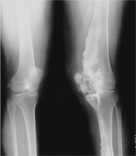 FIGURE 15-5 Melorheostosis. Standing views of the knees showing sclerosis and cortical thickening that crosses the joint into the soft tissues. The tibia also is involved. |
Suggested Reading
Morris JM, Samilson RL, Corey CL. Melorheostosis. J Bone Joint Surg 1963;45A:1191–1206.
Progressive Diaphyseal Dysplasia (Engelmann Disease)
Key Facts
Engelmann disease results in cortical thickening in the diaphysis of long bones progressing proximally and distally in the involved structure.
Most patients present in infancy or early childhood. It is an autosomal dominant inherited condition.
Neuromuscular dystrophy and malnutrition are associated with this condition.
Radiographic features:
Symmetric distribution
Diaphyseal cortical thickening involving endosteal and periosteal surfaces
Normal epiphysis and metaphysis
Relative elongation of involved extremities
Muscle atrophy
Differential diagnosis: chronic infection, infantile cortical hyperostosis, fibrous dysplasia
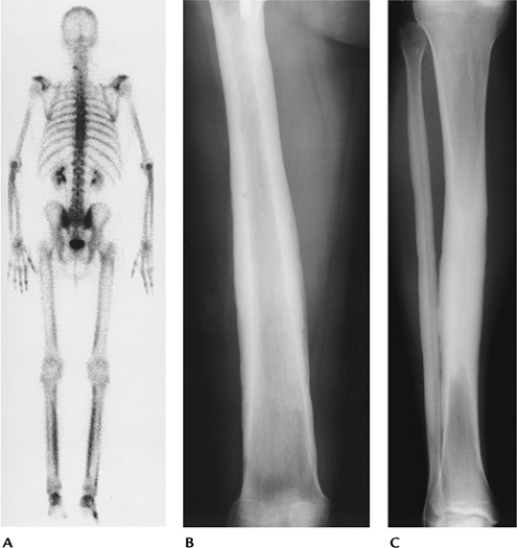 FIGURE 15-6 Engelmann disease. (A) Radionuclide bone scan showing symmetric increased cortical uptake in the femora, tibiae, and upper extremities. AP radiographs of the femur (B) and tibia (C) showing marked diaphyseal cortical thickening with sparing of the metaphyses and epiphyses. |
Suggested Reading
Kumar B, Murphy WA, Whyte MP. Progressive diaphyseal dysplasia (Engelmann disease): Scintigraphic-radiographic-clinical correlations. Radiology 1981;140:87–92.
Cleidocranial Dysplasia (Cleidocranial Dysostosis)
Key Facts
Cleidocranial dysplasia is an uncommon autosomal dominant disorder.
Patients present with delayed or incomplete cranial ossification and hypoplastic or aplastic clavicles. Delayed ossification may be evident in the axial skeleton and extremities.
The mandible may be large, and delayed tooth development is common.
Radiographic features:
Lack of midline ossification and wormian bones in the calvarium
Absent or hypoplastic clavicles
Delayed ossification in the spine, pelvis, and extremities
Femoral necks deformed or aplastic
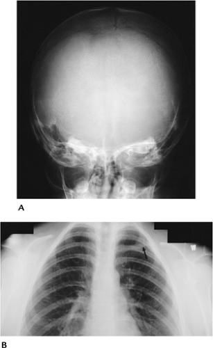 FIGURE 15-7 Cleidocranial dysplasia. (A) AP view of the skull showing multiple wormian bones along the suture lines. (B) AP view of the upper chest showing an absent right clavicle and small hypoplastic medial segment (arrow) on the left. |
Suggested Reading
Jarvis JL, Keats TE. Cleidocranial dysostosis. A review of 40 new cases. AJR Am J Roentgenol 1974;121:5–16.
Osteopetrosis
Key Facts
Osteopetrosis is a disease of uncertain cause that leads to dense brittle bones.
There are multiple clinical forms of this condition.
Osteopetrosis infantile: autosomal recessive with failure to thrive, hepatosplenomegaly, cranial nerve dysfunction, blindness, and deafness. Death frequently occurs in early years of life.
Osteopetrosis tarda (delayed): autosomal dominant. Patients usually are asymptomatic. Detection results from mild anemia, cranial nerve palsies, or pathologic fractures.
Osteopetrosis intermediate: autosomal recessive with features between infantile and tarda in severity.
Radiographic features:
Infantile: uniformly dense sclerotic bones with changes similar to rickets near the growth plates
Tarda: bone-within-a-bone appearance
Intermediate: diffuse bone sclerosis, especially of the skull base. Bone-within-a-bone appearance. Avascular necrosis of the femoral heads.
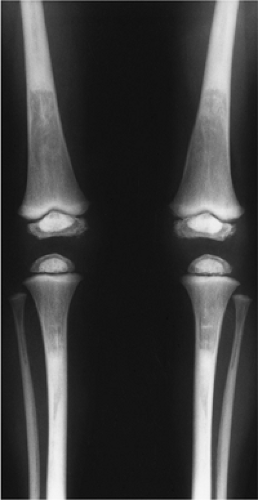 FIGURE 15-8 Osteopetrosis intermediate. AP radiographs of the tibia and femora showing bone sclerosis with bone-within-a-bone appearance in the epiphyses. |
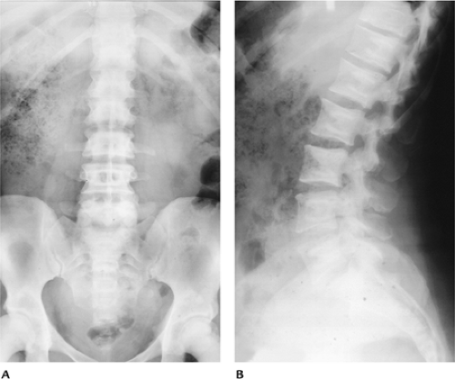 FIGURE 15-9 Osteopetrosis tarda. AP (A) and lateral (B) radiographs of the lumbar spine showing a bone-within-a-bone appearance. |
Suggested Reading
Shapiro F, Glimcher MJ, Holtrop ME, et al. Human osteopetrosis. J Bone Joint Surg 1980;62A:384–399.
Mastocytosis
Key Facts
Mastocytosis is a systemic disease with mast cell accumulation in multiple organs affecting adult males and females.
Liver, spleen, lymph node, skeletal, and most commonly cutaneous organs are involved.
Patients present with skin lesions resembling urticaria pigmentosa, also diarrhea, vomiting, flushing, or intermittent shocklike episodes.
Radiographic features occur in 70% of patients:
Osteopenia and bone destruction most common in the skull, spine, and ribs
Osteosclerosis, which may resemble metastasis, Paget disease, or myelofibrosis
Features may be focal or diffuse.
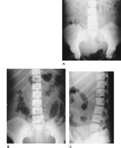 FIGURE 15-10 Mastocytosis. (A) AP radiograph of the lumbar spine and pelvis showing generalized bone sclerosis and cortical thickening. There are more focal foci of sclerosis in the femoral heads. AP (B) and lateral (C) radiographs of the lumbar spine and pelvis in a different patient showing diffuse small sclerotic foci. |
Suggested Reading
McKenna MJ, Frame B. The mast cell and bone. Clin Orthop 1985;200:226–233.
Tuberous Sclerosis
Key Facts
Tuberous sclerosis is an autosomal dominant inherited disorder.
Characteristic features include seizure disorders, mental retardation, and cutaneous hamartomas.
Radiographic features:
Skull: foci of sclerosis and trabecular prominence; calvarial thickening; intercerebral calcifications; brain lesions in ventricles, white matter, and cortex 50% to 80%.
Axial/appendicular skeleton: focal or diffuse cystlike lesions or areas of sclerosis. Subperiosteal and cortical lesions result in irregular cortical appearance.
Extraskeletal lesions: Fifty percent have renal cysts, angiolipomas, and aneurysms; 30% to 50% have rhabdomyomas of the heart. Pulmonary lesions in 1% commonly lead to pneumothorax.
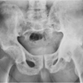
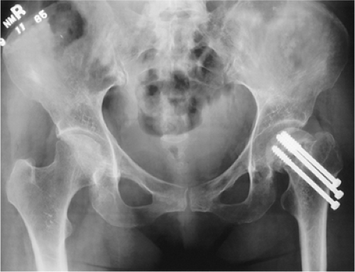 FIGURE 15-11 Tuberous sclerosis. AP radiograph of the pelvis showing oval- or flame-shaped areas of sclerosis in both iliac wings. There is an impacted left femoral neck fracture with pin fixation unrelated to the bone changes of tuberous sclerosis. |
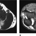
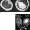
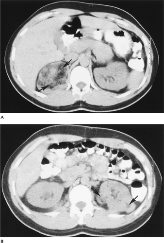 FIGURE 15-12 Tuberous sclerosis with renal angiomyolipomas. (A,B) Computed tomography (CT) images showing characteristic fat density masses (arrows), the largest in the right kidney. |
 FIGURE 15-13 Tuberous sclerosis. Axial T2-weighted MR image showing multiple areas of signal abnormality (arrows) resulting from cortical tubers. |
Suggested Reading
Medley BE, McLeod RA, Houser OW. Tuberous sclerosis. Semin Roentgenol 1976;11:35–54.
Wood B, Leiberman E, Larding B, et al. Tuberous sclerosis. AJR Am J Roentgenol 1992;158:750.
Neurofibromatosis
Key Facts
Neurofibromatosis is one of the most common inherited (autosomal dominant) disorders.
Clinical triad includes skin lesions, mental retardation, and skeletal deformities.
More than 99% of cases are in the category of neurofibromatosis Type 1 or Type 2.
• Type 1 (two or more features)
Six or more café-au-lait skin lesions
Two or more neurofibromas or one plexiform neurofibroma
Inguinal or axillary freckling
Optic glioma
Two or more iris hematomas
Osseous lesion
Parent, sibling, or child with Type 1
• Type 2 (one feature)
Bilateral eighth nerve masses
• Type 2 in parent, sibling, or child and an eighth nerve mass, or two of the following: neurofibroma, meningioma, glioma schwannoma, or posterior capsular lenticular capacity
Radiographic features:
Osseous features
Orbital and facial bone deformities
Spinal deformities (60%)
Scoliosis
Kyphoscoliosis
Vertebral scalloping
Pedicle erosion
Spindle ribs and transverse processes
Extremities
Bowing (especially tibia)
Pathologic fracture
Hypoplastic fibula
Pseudoarthrosis with pathologic fracture
Neural
Meningoceles
Cranial nerve tumors
Peripheral nerve neurofibromas and schwannomas
Malignant degeneration of neural lesions 2% to 29%
Other associated lesions
Neuroblastoma
Pheochromocytoma
Thyroid carcinoma
Wilms tumor
Rhabdomyosarcoma
Leukemia
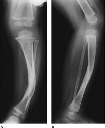 FIGURE 15-14 Neurofibromatosis Type 1. AP (A) and lateral (B) radiographs showing tibial bowing with a healed midtibial fracture. |
 FIGURE 15-15 Neurofibromatosis Type 1. Axial precontrast (A) and postcontrast (B) images showing enlargement of both optic nerves (arrows) caused by optic nerve gliomas. |
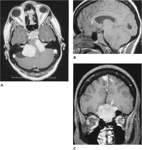 FIGURE 15-16 Neurofibromatosis Type 2. Bilateral vestibular nerve schwannomas and multiple meningiomas. (A) Postcontrast axial MR image showing bilateral large vestibular nerve schwannomas extending into the internal auditory canals and compressing the pons. Sagittal (B) and coronal enhanced (C) T1-weighted images showing multiple meningiomas (arrows). |
Suggested Reading
Sevick RJ, Barkovich AJ, Edwards MS, et al. Evolution of white matter lesions in neurofibromatosis type 1: MR findings. AJR Am J Roentgenol 1992;159:171–175.
Ollier Disease (Enchondromatosis)
Key Facts
Ollier disease is a noninherited condition resulting in multiple asymmetrically distributed enchondromas.
Lesions lead to fractures in adults and children.
In adults, lesions may undergo malignant degeneration to chondrosarcoma (5%–30%).
Radiographic features:
Multiple lytic expanding lesions are located predominantly in the extremities.
Flat bones of the pelvis also may be involved.
Lesions may contain calcification.
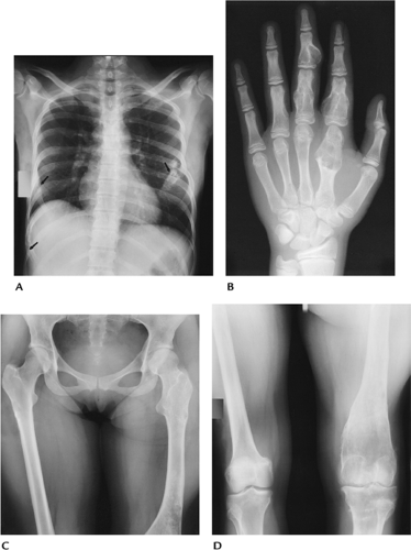 FIGURE 15-17 Ollier disease. (A) Posteroanterior (PA) chest radiographs showing multiple expanded calcified rib lesion (arrows). (B) PA view of the hand showing enchondromas in the second to fourth rays. AP radiographs of the pelvis (C) and femora (D) showing multiple enchondromas in the left femur. The largest expand the distal femur. |
Suggested Reading
Milgram JW. The origins of osteochondromas and enchondromas. A histopathologic study. Clin Orthop 1983;174:264–284.
Maffucci Syndrome
Key Facts
Maffucci syndrome is a rare disorder with multiple enchondromas and soft tissue hemangiomas.
The syndrome occurs in males and females, beginning in childhood.
Half of the cases are unilateral. The hand is most commonly involved.
Enchondromas may undergo malignant transformation to chondrosarcoma.
Radiographic features:
Multiple expanding lytic lesions that may contain calcifications
Soft tissue masses (hemangiomas) with phleboliths are characteristic.
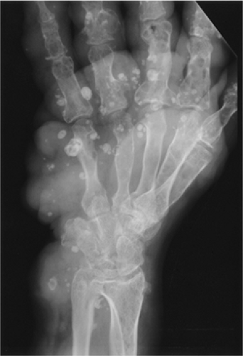 FIGURE 15-18 Maffucci syndrome. Oblique radiograph showing multiple enchondromas and soft tissue masses with vascular calcifications. |
Suggested Reading
Strang C, Ronnie I. Dyschondroplasia and hemangiomata (Maffucci’s syndrome). J Bone Joint Surg 1950;32B:376–383.
Hereditary Multiple Exostosis
Key Facts
Hereditary multiple exostosis is an autosomal dominant condition resulting in abnormal bone remodeling and bone deformities.
Patients present in childhood with palpable osseous masses, bone shortening, bowing, and joint deformities.
Osseous lesions relate to osteochondromas and are bilateral and near the physis.
Complications:
Pathologic fracture
Neurovascular injury
Bursa formation
Malignant degeneration (chondrosarcoma in 2% to 27%)
Radiographic features:
Osteochondroma-like lesions
Most common in the knee and proximal humerus
Lesions usually are bilateral and symmetric.
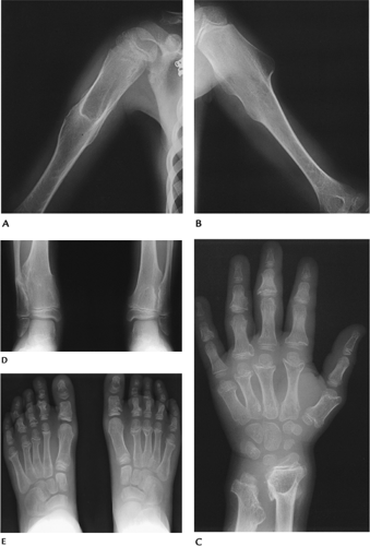 FIGURE 15-19 Hereditary multiple exostosis. Radiographs of the humeri (A,B), left hand and wrist (C), both ankles (D), and feet (E) demonstrate multiple exostoses with bone and joint deformities most obvious in the hand and wrist. |
Suggested Reading
Related posts:


Stay updated, free articles. Join our Telegram channel

Full access? Get Clinical Tree


