Fig. 27.1
A is the angle of necrotic area in mid-coronal image and B is the angle of necrotic area in midsagittal image. The angle is measured at the subchondral portion
The necrotic index was calculated by the formula: (A/180) × (B/180) × 100. In a pilot study to determine the reproducibility of this measurement, the coefficient of variation was 32 %. There was a strong correlation between this index and the risk of future collapse. If the cutoff point of 30 % is used, the predictive value of a negative test would be 100 % and that of a positive test 91 %. Likewise, a cutoff at 40 % gives a predictive value for a positive test of 100 % and for a negative test of 82 %. None of hips with necrosis less than 30 % collapsed, but all hips with more than 40 % necrosis collapsed, and half of hips with necrosis of between 30 and 40 % collapsed. Thus, hips with necrosis of less than 30 % are were classified as a low-risk group, those with necrosis of 30–40 % as a moderate-risk group, and those with more than 40 % as a high-risk group in terms of future collapse of the femoral head.
Although this method showed a strong correlation with the risk of collapse and the index was a major predictor of future collapse, a conversion table or a calculator was necessary to obtain the index.
27.2.2 The Second Modification
To simplify the method, a second modification was done in 2006 [14]. The arc of the necrotic portion on both the mid-coronal (A) and the midsagittal images (B) was measured (Fig. 27.1), and the sum of these two angles was then calculated. There was a strong correlation between the combined necrotic angle (A + B) and the risk of future collapse. None of hips with a combined necrotic angle of ≤190° collapsed, all hips with an angle of ≥240°collapsed, and 50 % of hips with an angle between 190° and 240° collapsed during the study period (Fig. 27.2). This would suggest that hips with a combined necrotic angle of ≤190° are in a low-risk group, those with an angle between 190° and 240° are in a moderate-risk group, and those with an angle of ≥240° are in a high-risk group.
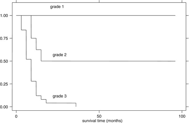
Fig. 27.2
Kaplan-Meier survival curve according to the categories of the combined necrotic angle, which were classified by cut points of 190° and 240°. Grade 1 denotes hips with combined necrotic angle ≤190°, grade 2 denotes hips with combined necrotic angle between 190° and 240°, and grade 3 denotes hips with combined necrotic angle ≥240°. Survival distributions for three grades were different by log rank test (p < 0.01)
27.3 Summary
The measurement of necrotic arc on mid-coronal and midsagittal MRI scans is simple and easy and reproducible. The combined necrotic angle, the sum of the two necrotic arcs on mid-coronal and midsagittal MRI scans, is based on biplanar MRI scans and provides a three-dimensional assessment of the extent of the necrosis. The categorization of femoral head osteonecrosis according to the combined necrotic angle may accurately predict the risk of subsequent collapse of the femoral head.
Medical or surgical treatment to prevent collapse is not justified in the low-risk group. Any intervention should be limited to hips in the moderate to high-risk groups.
References
1.
Related posts:
 Ischemia and Reperfusion Injury in Bone
 Glucocorticoids (as an Etiologic Factor)
 Alumina Ceramic-on-Highly Cross-Linked Polyethylene Bearing in Cementless Total Hip Arthroplasty in Young Patients with Osteonecrosis of Femoral Head
 Impaction Bone Grafting
 Causes of Pain in Early and Advanced Stage Osteonecrosis
 Kienböck’s Disease of the Lunate
Stay updated, free articles. Join our Telegram channel

Full access? Get Clinical Tree


