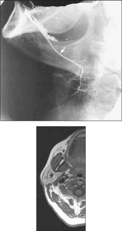
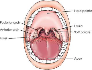
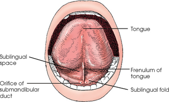
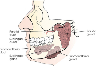
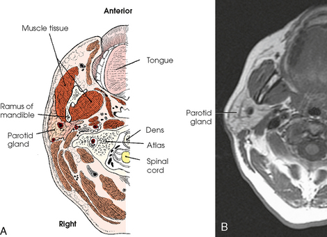
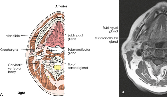
14 The mouth, or oral cavity, is the first division of the digestive system (Fig. 14-1). It encloses the dental arches and receives the saliva secreted by the salivary glands. The cavity of the mouth is divided into (1) the oral vestibule, the space between the teeth and the cheeks, and (2) the oral cavity, or mouth proper, the space within the dental arches. The roof of the oral cavity is formed by the hard and soft palates. The floor is formed principally by the tongue, and it communicates with the pharynx posteriorly via the oropharynx. The tongue is situated in the floor of the oral cavity, with its base directed posteriorly and its apex directed anteriorly (Fig. 14-2; see Fig. 14-1). The tongue is freely movable. The tongue is composed of numerous muscles and is covered with a mucous membrane that varies in complexity in the different regions of the organ. The extrinsic muscles of the tongue form the greater part of the oral floor. The mucous membrane covering the undersurface of the tongue is reflected laterally over the remainder of the floor to the gums. This part of the floor lies under the free anterior and lateral portions of the tongue and is called the sublingual space. Posterior movement of the free anterior part of the tongue is restricted by a median vertical band, or fold, of mucous membrane called the frenulum of the tongue, which extends between the undersurface of the tongue and the sublingual space. On each side of the frenulum, extending around the outer limits of the sublingual space and over the underlying salivary glands, the mucous membrane is elevated into a crestlike ridge called the sublingual fold. In the relaxed state, the two folds are quite prominent and are in contact with the gums. The three pairs of salivary glands produce approximately 1 L of saliva each day. The glands are named the parotid,! submandibular, and sublingual (Fig. 14-3). Each gland is composed of numerous lobes, and each lobe contains small lobules. The whole gland is held together by connective tissue and a fine network of blood vessels and ducts. The minute ducts of the lobules merge into larger tributaries, which unite and form the large efferent duct that conveys the saliva from the gland to the mouth. The parotid glands, the largest of the salivary glands, each consist of a flattened superficial portion and a wedge-shaped deep portion (Fig. 14-4). The superficial part lies immediately anterior to the external ear and extends inferiorly to the mandibular ramus and posteriorly to the mastoid process. The deep, or retromandibular, portion extends medially toward the pharynx. The parotid duct runs anteriorly and medially to open into the oral vestibule opposite the second upper molar. The submandibular glands are large, irregularly shaped glands. On each side, a submandibular gland extends posteriorly from a point below the first molar almost to the angle of the mandible (Fig. 14-5). Although the upper part of the gland rests against the inner surface of the mandibular body, its greater portion projects below the mandible. The submandibular duct extends anteriorly and superiorly to open into the mouth on a small papilla at the side of the frenulum of the tongue. The sublingual glands, the smallest pair, are narrow and elongated in form (see Fig. 14-5). These glands are located in the floor of the mouth beneath the sublingual fold. Each is in contact with the mandible laterally and extends posteriorly from the side of the frenulum of the tongue to the submandibular gland. Numerous small sublingual ducts exist. Some of these ducts open into the floor of the mouth along the crest of the sublingual fold, and others open into the submandibular duct. The main sublingual duct opens beside the orifice of the submandibular duct.
MOUTH AND SALIVARY GLANDS
Mouth
Salivary Glands

Related posts:
Stay updated, free articles. Join our Telegram channel

Full access? Get Clinical Tree


