© Springer-Verlag Berlin Heidelberg 2014
Ralf Puls and Norbert Hosten (eds.)Whole-body MRI Screening10.1007/978-3-642-55201-4_22. MR Imaging in Population-Based Research
(1)
Institute for Community Medicine, Study of Health in Pomerania (SHIP)/Clinical-Epidemiological Research, University Medicine Greifswald, Walther-Rathenau-Straße 48, Greifswald, 17487, Germany
2.1 Introduction
Epidemiologic research is aimed at estimating the prevalence and incidence of risk factors and diseases and at identifying the complex associations that exist among them. A central approach used by epidemiologists is to observe the evolution of both risk factors and diseases over time.
In a classic cohort study, investigators compile baseline data on risk factors pertinent to the questions they seek to answer and relate them to the later occurrence of diseases and to mortality. A case in point is the Framingham Heart Study, which began in 1948 and provided the data that led to the identification of an association between serum lipid levels and the risk of developing coronary heart disease later in life (Kannel et al. 1964).
With the technical advances made over the last decades, imaging modalities have gained an increasingly important role in clinical routine and in epidemiologic research. In epidemiologic studies, imaging examinations are performed to diagnose clinical diseases and identify subclinical conditions. The methods used include photography, ultrasound examinations, and MR and CT imaging.
2.1.1 Diagnosis of Clinical Disease
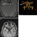
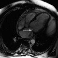
Health-related data obtained by interview or questionnaire often have limited validity. Both acute and chronic conditions may be asymptomatic, or signs and symptoms are overlooked because they are mild or because an individual’s perception is reduced or distorted. In such cases, imaging modalities provide objective and independent evidence of prior or existing diseases. For instance, MR imaging will detect brain lesions persisting after a stroke or a myocardial scar secondary to a silent myocardial infarction.
2.1.2 Detection of Subclinical Abnormalities
Subclinical changes on MR images are not diseases but may point to beginning or ongoing disease processes that would otherwise go unnoticed. In the development of diseases, such subclinical changes are intermediate between risk factors and outcome, which is why they are often considered to have a high predictive value for the development of clinical disease and are therefore of interest as surrogate parameters.
Imaging can detect morphologic and functional subclinical changes. Morphologic changes are apparent on individual images, while dysfunction requires a series of images, obtained in some cases after provocation. Examples of relevant subclinical alterations include an increase in carotid wall thickness (Chambless et al. 2004) and reduced flow-mediated vasodilation (Shimbo et al. 2007). These can be detected by ultrasound and are considered to be indicators of an increased risk of atherosclerosis. Carotid wall thickness is measured with the subject at rest and is a morphologic finding, while the degree of loss of flow-mediated vasodilation is a functional finding and is estimated during a brief high-flow state in the brachial artery following temporary ischemia.
2.2 Imaging in the Study of Health in Pomerania
The Study of Health in Pomerania (SHIP) is a population-based study conducted in the northeast of Germany (Völzke et al. 2011). Its goal is to estimate the prevalence and incidence of risk factors and clinical diseases in the general population of the German Federal State of Mecklenburg-Vorpommern, elucidating the complex relationships among risk factors, subclinical findings, and clinical diseases. A distinctive feature of SHIP is that it does not focus on a specific organ or organ system but instead aims at characterizing health and disease in a very comprehensive way.
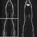
SHIP investigators have been using imaging modalities for detecting subclinical abnormalities since 1997 when the study began. In the baseline examination (SHIP-0), ultrasound was used to measure carotid wall thickness and to detect atherosclerotic plaque; to determine thyroid size and to identify thyroid nodules; to evaluate cardiac valve function, left ventricular mass, and pump function; and to diagnose fatty liver and gallstones. Additional imaging modalities were introduced for 5-year follow-up examinations (SHIP-1). These included measurement of flow-mediated vasodilation as well as photography and sonographic examinations to identify varicose veins below the knee.
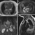
The spectrum of imaging modalities was expanded even further for the most recent investigations – the 11-year follow-up study of the first SHIP cohort (SHIP-2) and the baseline examinations of a second, independent SHIP cohort (SHIP-Trend). The additional procedures include photography to document and quantify changes in the ocular fundus and ultrasound to estimate bone stiffness and to identify morphologic changes in the kidneys and pancreas.
MR imaging is staff-intensive and expensive, which is why it is rarely used as an imaging tool in population-based research. All current studies making use of this imaging modality do so for dedicated examinations of individual organs or body regions. Examples include MR brain scans in the Rotterdam Study (Vernooij et al. 2007), cardiac MR imaging in the Multi-Ethnic Study of Atherosclerosis (Nasir et al. 2007), and MR spectroscopy for quantifying hepatic fat content in the Dallas Heart Study (Romeo et al. 2008). SHIP-2 and SHIP-Trend are the first population-based studies using whole-body MR imaging on a large scale (Hegenscheid et al. 2009). The use of whole-body MR imaging in epidemiology has considerable scientific potential but also presents great challenges in terms of quality assurance and application of ethical standards.
2.3 Scientific Potential of Whole-Body MR Imaging Examinations in Population-Based Research
The large-scale use of whole-body MR imaging in a population-based cross-sectional study provides a wealth of data for answering a multitude of scientific questions of high clinical relevance. These include the establishment of reference values for MR examinations, prevalence estimates, and association studies.
Related posts:
Stay updated, free articles. Join our Telegram channel

Full access? Get Clinical Tree


