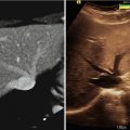
Fig. 43.1
Preoperative axial MRI with contrast demonstrating a metastatic brainstem tumor that has enlarged over time. The cerebellar tumors have been stable
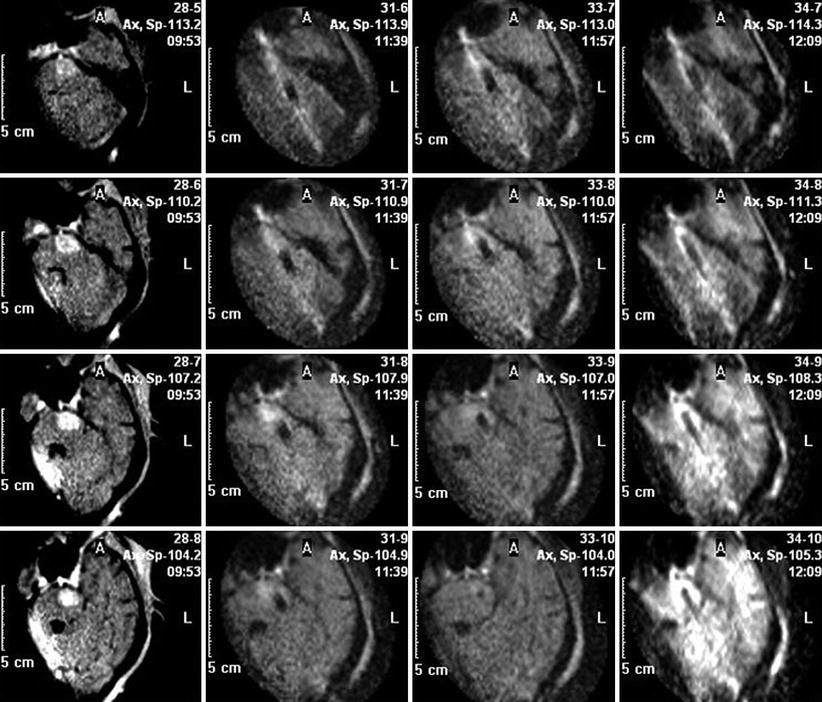
Fig. 43.2
Intraoperative MRI compare function, showing images over time, from left to right. Left, preoperative scan showing brainstem metastasis. Following images demonstrate the cannula for laser treatment being positioned within the tumor. Right column shows the laser fiber itself within the lesion
The patient was then transferred to a 1.5 T MRI for LITT. Tumor ablation maps from the procedure are shown in Fig. 43.3. To complete tumor lysis, the laser fiber was progressively withdrawn until 3 LITT treatments were done. MRI immediately after LITT showed an absence of contrast enhancement, consistent with tumor ablation (Fig. 43.4). The patient initially had a slight increase in her ataxia after surgery but recovered within several weeks. She died of progressive systemic disease 10 months after LITT.
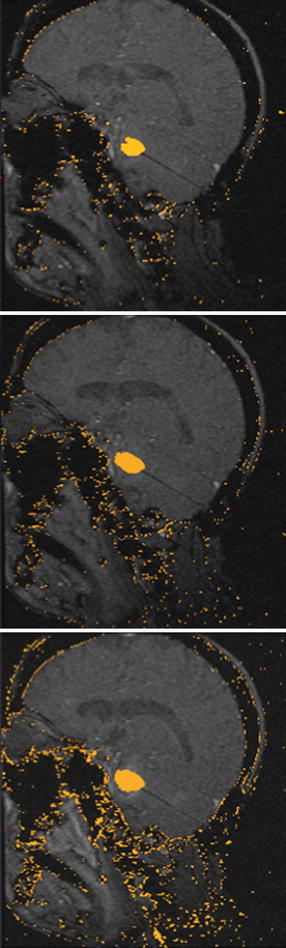
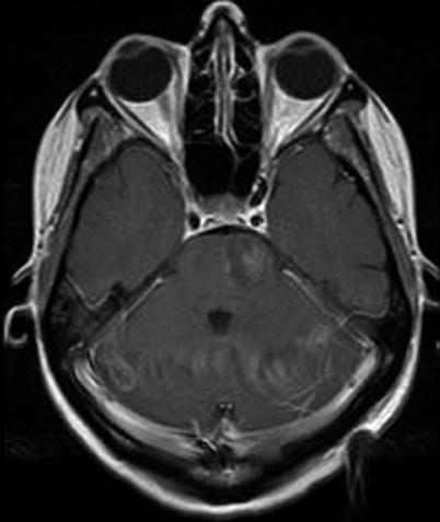
Fig. 43.3
Tumor ablation maps during LITT of the brainstem metastasis. The lesion is “grown” by withdrawing the laser fiber and repeating heat delivery to 60 °C
Fig. 43.4
MRI after LITT shows the absence of contrast enhancement, consistent with ablation of the tumor
Our Experience
We have used LITT to treat six patients with intracranial tumors and one with a recurrent ependymoma of the conus region (see Table 43.1). In all cases, the patient was seeking alternative opinions after multiple surgical resections had been attempted and lesion recurrence observed after multiple chemotherapy and radiation therapies tried. Nonetheless, most patients referred to our protocol included those with multiple intracranial metastases. However, patients with other recurrent CNS tumors have been referred as well (Table 43.2). Intraoperative MRI was used for surgical navigation and fiber placement confirmation in 5/7 patients (including the lone spinal tumor patient). In 3/5 cases, iMRI was used to improve laser placement. This includes one patient in whom an initial attempt with surgical navigation alone to place two lasers to treat a right parietal metastasis had to be aborted after the lasers were off target when imaged at 1.5 T. The repeat procedure with iMRI proved successful.
Table 43.1
Patient characteristics
Age (years) | |
Median | 63 |
Range | 39–72 |
Sex | |
Male | 2 |
Female | 5 |
Prior treatments (no. patients) | |
Surgery | 13 (5) |
Radiation | 9 (6)
Related posts:Stay updated, free articles. Join our Telegram channel
Full access? Get Clinical Tree
 Get Clinical Tree app for offline access

|